. 2021 Aug;36(2):289-296.
doi: 10.1177/0885328221999894. Epub 2021 Mar 12.

Emulsion-free chitosan-genipin microgels for growth plate cartilage regeneration

Affiliations

Emulsion-free chitosan-genipin microgels for growth plate cartilage regeneration

Christopher Erickson et al. J Biomater Appl. 2021 Aug.

Abstract

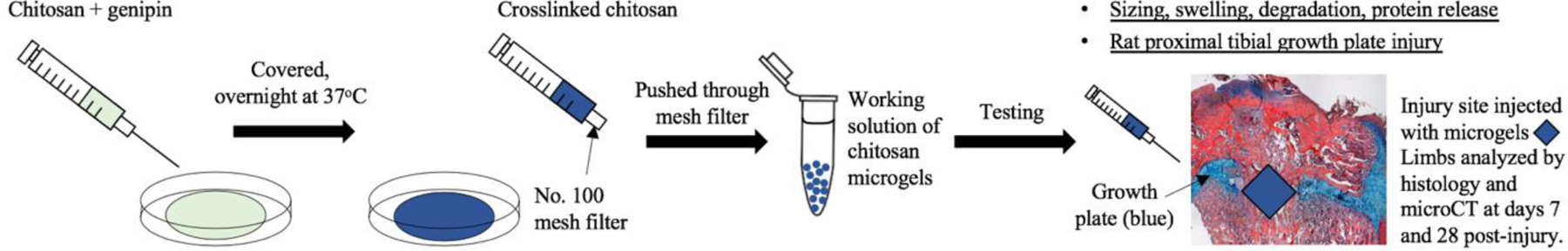
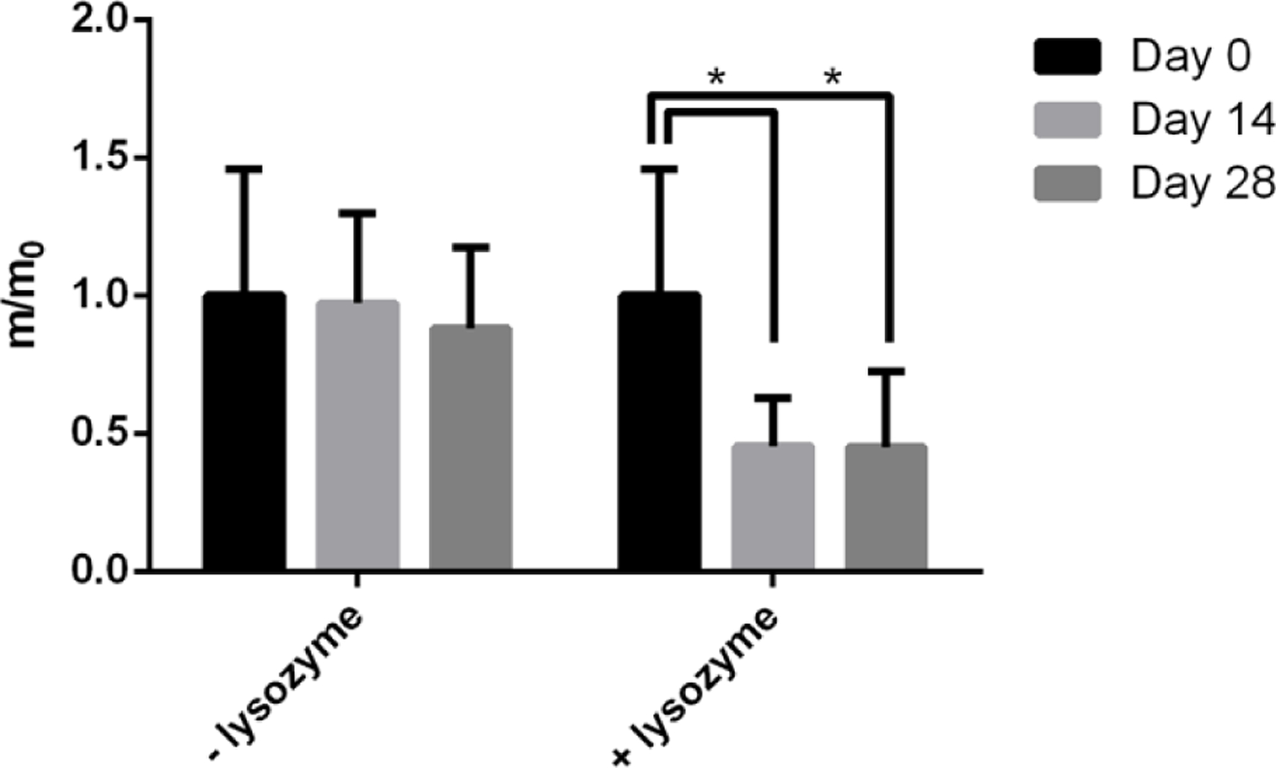
The growth plate is a cartilage tissue near the ends of children's long bones and is responsible for bone growth. Injury to the growth plate can result in the formation of a 'bony bar' which can span the growth plate and result in bone growth abnormalities in children. Biomaterials such as chitosan microgels could be a potential treatment for growth plate injuries due to their chondrogenic properties, which can be enhanced through loading with biologics. They are commonly fabricated via an emulsion method, which involves solvent rinses that are cytotoxic. Here, we present a high throughput, non-cytotoxic, non-emulsion-based method to fabricate chitosan-genipin microgels. Chitosan was crosslinked with genipin to form a hydrogel network, and then pressed through a syringe filter using mesh with various pore sizes to produce a range of microgel particle sizes. The microgels were then loaded with chemokines and growth factors and their release was studied in vitro. To assess the applicability of the microgels for growth plate cartilage regeneration, they were injected into a rat growth plate injury. They led to increased cartilage repair tissue and were fully degraded by 28 days in vivo. This work demonstrates that chitosan microgels can be fabricated without solvent rinses and demonstrates their potential for the treatment of growth plate injuries.

Keywords: Chitosan; cartilage regeneration; growth plate; injectable biomaterials; microgels.


Figures

Figure 1:
Figure 1:
Schematic of study.
Figure 2:
Figure 2:
(a) Normal distribution graph of the Feret Diameter showing swelling behavior of microgels in response to pH changes. (b) Fluorescent images of microgels fabricated using No. 200 mesh (upper image. <75 µm sized microgels), and No. 100 mesh (lower image. ~75–150 µm sized microgels).
Figure 3:
Figure 3:
Microgel degradation in the presence of lysozyme in 20 mM HEPES pH 6.8 buffer at Day 0, Day 14 and Day 28. * p<0.05 compared to the Day 0 time point.
Figure 4:
Figure 4:
Release curves showing the cumulative protein release over time of SDF-1α (a) and TGF-β3 (b) from microgels.
Figure 5:
Figure 5:
10x histological images showing growth plate repair tissue of intact (a, e), untreated (b, f), microgel treated (c, g), microgel + SDF-1α treated (d, h), and microgel + TGF-β3 (i) limbs. No day 7 animals were treated with microgel + TGF-β3. ABH stains the bone orange to red, fibrous tissue pink, and cartilage blue. The microgel appears as a dark red fibrous-like tissue. Scale bars = 500 µm.
Figure 6:
Figure 6:
Graphs showing bony bar formation at 7 (a) and 28 (b) days post-injury within the growth plate injury site as determined by microCT. (c–k) are microCT images of the tibiae, showing the growth plate and bony bar formation in response to the different treatments at both time points. The growth plate is the thin, radiolucent tissue (c, g). * indicates statistical significance (p < 0.05) versus the untreated group within the day 7 time point. The yellow box in (d) indicates how the ROI was drawn to measure bone growth within the growth plate injury. N =3–4 animals per group.

References

    1. Pella MCG, Lima-Tenorio MK, Tenorio-Neto ET, et al. Chitosan-based hydrogels: From preparation to biomedical applications. Carbohydr Polym 2018; 196: 233–245. 2018/06/13. DOI: 10.1016/j.carbpol.2018.05.033. - DOI - PubMed
    1. Felt O, Buri P and Gurny R. Chitosan: a unique polysaccharide for drug delivery. Drug development and industrial pharmacy 1998; 24: 979–993. 1999/01/07. DOI: 10.3109/03639049809089942. - DOI - PubMed
    1. Karnchanajindanun J, Srisa-ard M and Baimark Y. Genipin-cross-linked chitosan microspheres prepared by a water-in-oil emulsion solvent diffusion method for protein delivery. Carbohydrate Polymers 2011; 85: 674–680. DOI: 10.1016/j.carbpol.2011.03.035. - DOI
    1. Yan L-P, Wang Y-J, Ren L, et al. Genipin-cross-linked collagen/chitosan biomimetic scaffolds for articular cartilage tissue engineering applications. Journal of Biomedical Materials Research Part A 2010; 95A: 465–475. DOI: 10.1002/jbm.a.32869. - DOI - PubMed
    1. Muzzarelli RA, El Mehtedi M, Bottegoni C, et al. Genipin-Crosslinked Chitosan Gels and Scaffolds for Tissue Engineering and Regeneration of Cartilage and Bone. Mar Drugs 2015; 13: 7314–7338. 2015/12/23. DOI: 10.3390/md13127068. - DOI - PMC - PubMed

Publication types

LinkOut - more resources