. 2021 Mar 12;58(2):634-645.
doi: 10.1093/jme/tjaa247.

Morphological Characterization of the Antennal Sensilla of the Afrotropical Sand Fly, Phlebotomus duboscqi (Diptera: Psychodidae)

Affiliations

Morphological Characterization of the Antennal Sensilla of the Afrotropical Sand Fly, Phlebotomus duboscqi (Diptera: Psychodidae)

Ana Cristina Bahia et al. J Med Entomol. .

Erratum in

Abstract

We investigated by scanning electron microscopy the morphology, distribution, and abundance of antennal sensilla of females Phlebotomus duboscqi sand fly, an important vector of zoonotic cutaneous leishmaniasis at Afrotropical region. Thirteen well-differentiated sensilla were identified, among six types of cuticular sensilla. The probable function of these sensillary types is discussed in relation to their external structure and distribution. Five sensillary types were classified as olfactory sensilla, as they have specific morphological characters of sensilla with this function. Number and distribution of sensilla significantly differed between antennal segments. The results of the present work, besides corroborating in the expansion of the morphological and ultrastructural knowledge of P. duboscqi, can foment future electrophysiological studies for the development of volatile semiochemicals, to be used as attractants in traps for monitoring and selective vector control of this sand fly.

Keywords: Leishmania major; Phlebotominae; antenna; sand fly; ultrastructure.


Figures

Fig. 1.
Fig. 1.
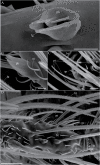
SEM of the head of females Phlebotomus duboscqi. (A) Partial frontal view of the head showing the compound eyes, with numerous ommatidia (om) and, among them, the insertion of the antenna, the scapus (S), the pedicellus (P), and proximal part of the first segment of the flagellum (I flg), showing to be covered by countless smaller noninnervated spinules of microtrichia type (m). Short sharp-tipped (st) sensilla were observed in the more proximal part of S and P, arranged in a set of three on both segment, as well as squamiform (sq) sensilla. In the median part of S, P, and first flagellomere (I flg), long blunt-tipped (bt) sensilla were found. In the most distal part of the pedicellus, campaniform sensilla (c) was evidenced (circle area) (scale bar = 25.3 µm). (B) Higher magnification of the short sharp-tipped (st) sensilla, and microtrichia (m), found on the scapus, and on the pedicellus (C) (scale bars = 5 µm, both). (D) Higher magnification of the circle area of scapus showing a pair of campaniform (c) sensilla, and microtrichia (m; scale bar = 2.6 µm). (E) Microtrichiae (m), the insertion (is) of a long blunt-tipped (bt) sensillum, and the serrated aspect of the surface of this sensillum, formed by the linear row of juxtaposed spines, were observed (scale bar = 2 µm). (F) A similar aspect is observed on the dorsal face (d) of squamiform (sq) sensilla, which has an almost smooth ventral face (v), with only a few longitudinal prominences in its proximal part (scale bar = 2.8 µm). (G) Ventral view of the scapus (S) showing a pair of short sharp-tipped (st) sensilla implanted proximally, and ommatidia (om; scale bar = 10 µm).
Fig. 2.
Fig. 2.
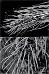
SEM of the antennae of females Phebotomus duboscqi. (A) Dorsolateral view of part of the pedicellus (P) and the first flagellomere (I flag) showing long blunt-tipped (bt) and squamiform (sq) sensilla (s), besides a set of more delicate sensilla (circle area; scale bar = 10 µm). (B) Higher magnification of circle area showing microtrichia (m), two short blunt-tipped (sbt) sensilla close to a set of six basiconic (b) sensilla (scale bar = 6 µm). (C) Higher magnification of short blunt-tipped (sbt) sensilla (scale bar = 1.1 µm), and (D) basiconic sensilla (scale bar: 1. 2 µm). (E) Flagellomere VI showing a pair of chaetic (ch) sensilla implanted, symmetrically arranged, besides in long blunt-tipped (bt) (scale bar = 20 µm). (F) Proximal part of the chaetic (ch) sensilla showing its implantation base (scale bar = 4.2 µm). (G) Higher magnification of the surface of this sensilla showing the presence of wall pores (scale bar = 0.43 µm).
Fig. 3.
Fig. 3.
SEM of the flagellum of females Phebotomus duboscqi. (A) Flagellomere III showing praying hands coeloconic (ph) sensillum with inner cone presenting longitudinal wall groves (lg; scale bar = 2 µm). (B) Lateral view of praying hands coeloconic (ph) sensillum, long blunt-tipped (bt), and microtrichia (m; scale bar = 1 µm). (C) Flagellomere XIII showing long blunt-tipped (bt), medium pointed-tipped (pt), common grooved coeloconic (gc), and short prominent-base (sp) sensilla (scale bar = 2 µm). (D) Flagellomere XII showing long blunt-tipped (bt), medium pointed-tipped (pt), common grooved coeloconic (gc), and chaetic (ch) sensilla (scale bar = 10 µm).
Fig. 4.
Fig. 4.
SEM of the flagellum of females Phebotomus duboscqi. (A) Flagellomere XIII showing long blunt-tipped (bt), common grooved coeloconic (gc), chaetic (ch), and medium pointed-tipped (pt) sensilla (scale bar = 10 µm). (B) Flagellomere X showing chaetic (ch), medium pointed-tipped (pt), long blunt-tipped (bt), and microtrichiae (m; scale bar = 5 µm). (C) Flagellomere IV showing common grooved coeloconic sensillum (gc), containing an inner cone with longitudinal wall grooves (lg) in its wall, surrounded by microtrichiae (m), besides long blunt-tipped (bt) and medium pointed-tipped (pt) sensilla (scale bar = 2 µm).
Fig. 5.
Fig. 5.
SEM of the flagellum of females Phebotomus duboscqi. (A) Flagellomere XIV showing common grooved coeloconic (gc), medium pointed-tipped (pt), short fixed-base sensilla (sf), apical trichoid (ap), and long blunt-tipped (bt) sensilla (scale bar = 10 µm). (B) Distal part of the last antennal flagellomere showing one apical trichoid (ap) sensillum, on its apex, short fixed-base (sf), medium pointed-tipped (pt), and common grooved coeloconic (gc) sensilla (scale bar = 5 µm). (C) Higher magnification of short fixed-base sensilla (sf; scale bar = 3 µm).
Fig. 6.
Fig. 6.
Size and number of the antennal segments of Phebotomus duboscqi females and of some of their antennal sensilla (P < 0.05). (A) Size of the antennal segments. (B) Length of the single-base chaetic sensilla, observed only in the antennal subsegments I–XIII. (C) Total number of sensilla on the antennal segments. (D) Number of long blunt-tipped sensilla found on antennal segments. (E) Number of medium pointed-tipped sensilla found on the last three antennal subsegments. (F) Number of common grooved coeloconic sensilla found on the last three antennal subsegments.

References

    1. Altner, H., and Prillinger L.. 1980. Ultrastructure of invertebrate chemo-, thermo-, and hygroreceptors and its functional significance. Int. Rev. Cytol. 67: 69–139.
    1. (AFPMB) Armed Forces Pest Management Board . 2015. Sand flies – significance, surveillance, and control in contingency operations. Technical Guide No. 49. Armed Forces Pest Management Board, US Army Garrison, Silver Spring, MD. 65 p.
    1. Bahia, A. C., Secundino N. F., Miranda J. C., Prates D. B., Souza A. P., Fernandes F. F., Barral A., and Pimenta P. F.. 2007. Ultrastructural comparison of external morphology of immature stages of Lutzomyia (Nyssomyia) intermedia and Lutzomyia (Nyssomyia) whitmani (Diptera: Psychodidae), vectors of cutaneous leishmaniasis, by scanning electron microscopy. J. Med. Entomol. 44: 903–914. - PubMed
    1. Bray, D. P., Ward R. D., James A., and Hamilton G. C.. 2010. The chemical ecology of sandflies, pp. 203–215. InTakken W. and Knols B. G. J.. (eds.), Olfaction in vector-host interactions: ecology and control of vector-borne diseases, vol. 2. Wageningen Academic Publishers, Wageningen, The Netherlands.
    1. Cate, H. S., and Derby C. D.. 2002. Hooded sensilla homologues: structural variations of a widely distributed bimodal chemomechanosensillum. J. Comp. Neurol. 444: 345–357. - PubMed

Publication types