. 2021 Apr 9;7(4):746-758.
doi: 10.1021/acsinfecdis.0c00437. Epub 2021 Mar 12.

A Long Residence Time Enoyl-Reductase Inhibitor Explores an Extended Binding Region with Isoenzyme-Dependent Tautomer Adaptation and Differential Substrate-Binding Loop Closure

Affiliations

A Long Residence Time Enoyl-Reductase Inhibitor Explores an Extended Binding Region with Isoenzyme-Dependent Tautomer Adaptation and Differential Substrate-Binding Loop Closure

Sandra Eltschkner et al. ACS Infect Dis. .

Abstract

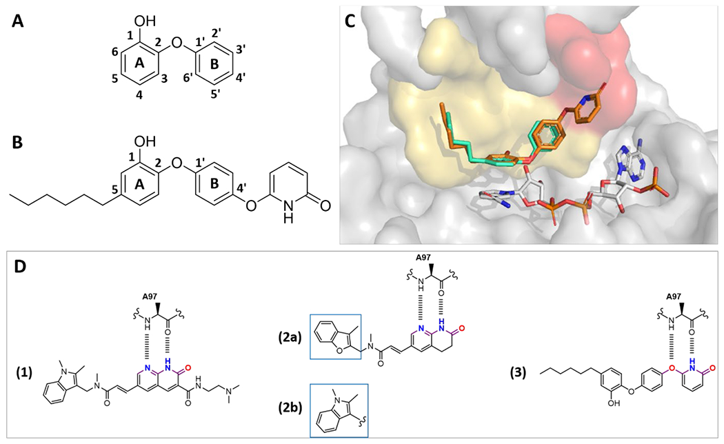
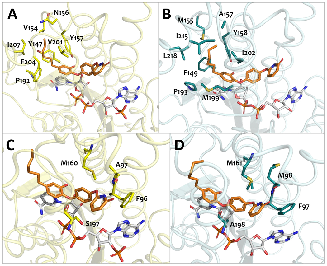
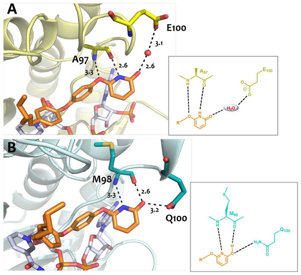
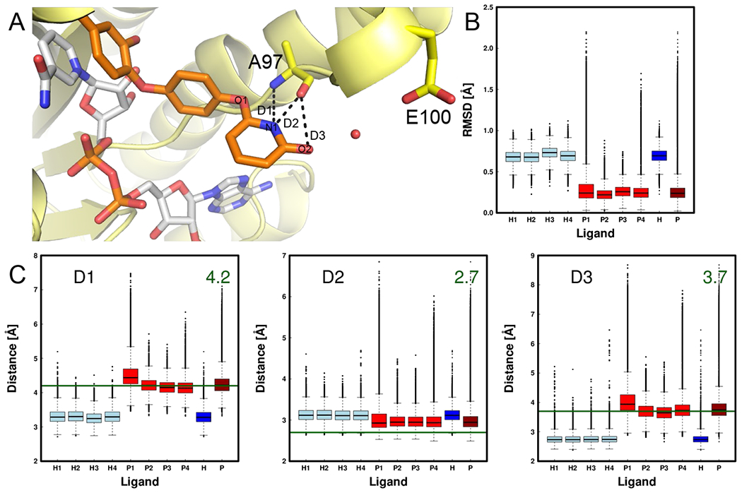
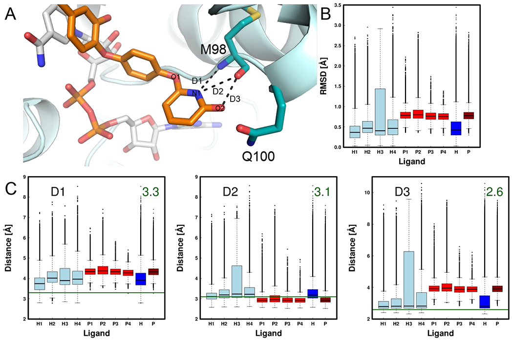
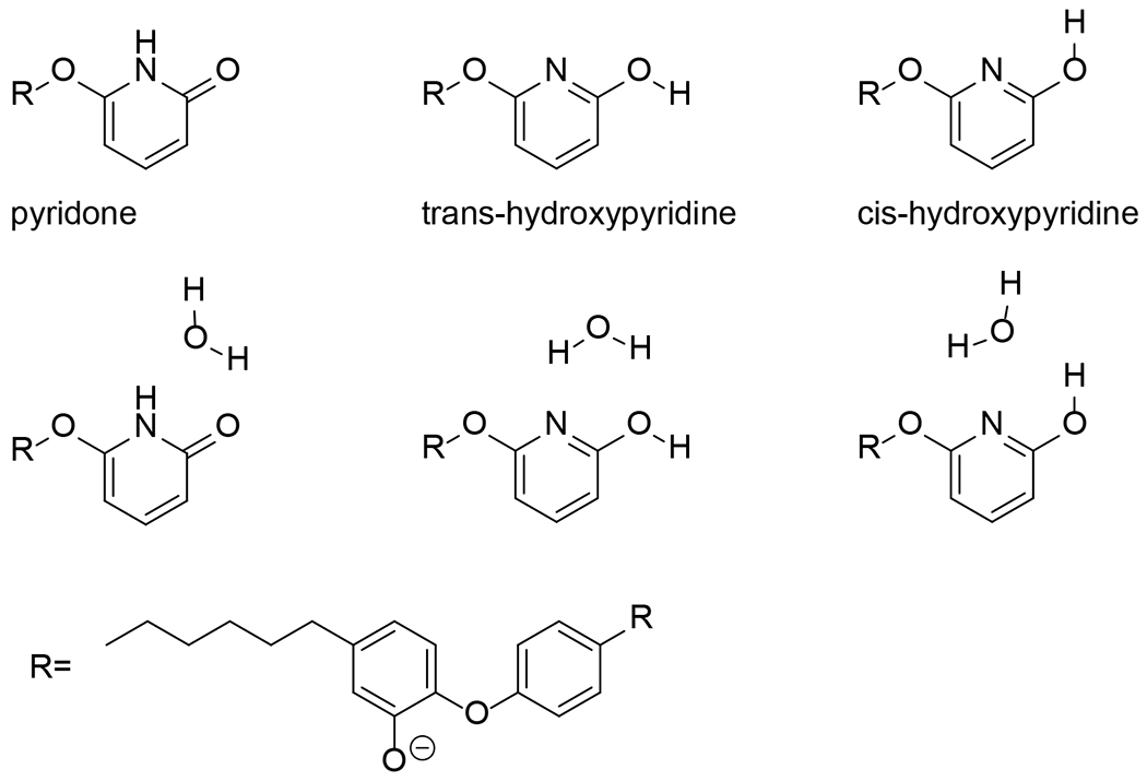
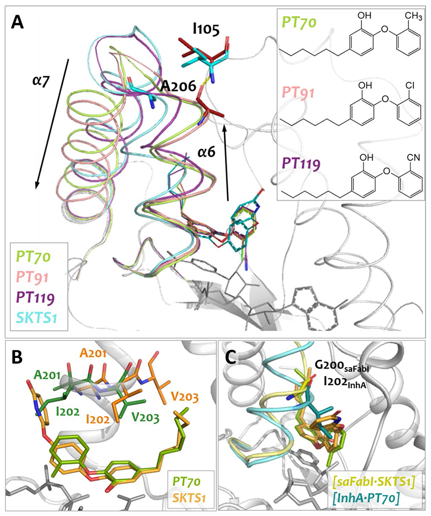
The enoyl-acyl carrier protein (ACP) reductase (ENR) is a key enzyme within the bacterial fatty-acid synthesis pathway. It has been demonstrated that small-molecule inhibitors carrying the diphenylether (DPE) scaffold bear a great potential for the development of highly specific and effective drugs against this enzyme class. Interestingly, different substitution patterns of the DPE scaffold have been shown to lead to varying effects on the kinetic and thermodynamic behavior toward ENRs from different organisms. Here, we investigated the effect of a 4'-pyridone substituent in the context of the slow tight-binding inhibitor SKTS1 on the inhibition of the Staphylococcus aureus enoyl-ACP-reductase saFabI and the closely related isoenzyme from Mycobacterium tuberculosis, InhA, and explored a new interaction site of DPE inhibitors within the substrate-binding pocket. Using high-resolution crystal structures of both complexes in combination with molecular dynamics (MD) simulations, kinetic measurements, and quantum mechanical (QM) calculations, we provide evidence that the 4'-pyridone substituent adopts different tautomeric forms when bound to the two ENRs. We furthermore elucidate the structural determinants leading to significant differences in the residence time of SKTS1 on both enzymes.

Keywords: Mycobacterium tuberculosis; Staphylococcus aureus; diphenylether; enoyl-ACP reductase; residence time; tautomerization.


Figures

Figure 1:
Figure 1:. The diphenylether (DPE) scaffold, substitution sites and novel target area of the 4’-pyridone within the substrate-binding pocket.
A) Ring A and B of the DPE scaffold are connected via an ether bridge. B) The DPE derivative SKTS1 contains a hexyl chain at position 5 and a pyridone substituent at the 4’-position. C) Comparison of the substrate-binding pocket surface in saFabI opposite of the substrate-binding loop (SBL) that is addressed by PT03 (greencyan, PDB: 4BNG, monomer A) and in our crystal structure with SKTS1 (orange, PDB code: 6YUR). The common contact surface of DPEs is shown in yellow; the extended area explored by the SKTS1 4’-pyridone is coloured red. The SBL is omitted for clarity. PT03, SKTS1 and NADP+ are shown as sticks. D) The naphthyridinone moieties of CG400462 (1), AFN-1252 (2a) and a derivative (2b) address the backbone atoms of A97 in the sa/bcFabI substrate-binding pocket and served as templates for the addition of an isosteric moiety to the DPE scaffold of SKTS1 (3). The isosteric portion of the inhibitors is coloured in magenta with oxygen and nitrogen atoms (with corresponding hydrogens) shown in red and blue, respectively.
Figure 2:
Figure 2:. Major interactions of SKTS1 with saFabI and InhA.
A, B) Hydrophobic interactions of SKTS1 in the saFabI (A, monomer A) and InhA (B) substrate-binding pockets. The residues’ main-chain atoms are hidden for clarity. C, D) Interactions of the B ring and the 4’-pyridone/hydroxypyridine substituent with saFabI (C) and InhA (D). The cofactor (grey), inhibitor (orange) and interacting residues (saFabI: yellow, InhA: deepteal) are shown as sticks; the overall protein scaffold is depicted in cartoon representation.
Figure 3:
Figure 3:. Interactions of the 4’-pyridone/hydroxypyridine ring with the protein environment.
A) Hydrogen bonds of the pyridone tautomer with saFabI. B) Hydrogen bonds of the hydroxypyridine tautomer with InhA. The putative protonation states are shown as a schematic view in the insets. Hydrogen bonds are shown as black dashed lines with distances given in Å.
Figure 4:
Figure 4:. RMSD- and distance-based comparison of the two tautomers for simulations of SKTS1 bound to saFabI.
A) Binding pose in the crystal structure. The ligand is shown in orange, NADP+ in white and the protein in yellow. Important distances D1, D2 and D3 are labelled accordingly. B) RMSD value of the N and O backbone atoms of A97 together with the N and O atoms of the pyridone/hydroxypyridine moiety after alignment of the trajectory to these atoms of the respective monomer in the crystal structure. The values for the individual ligands in each monomer (numbered 1 to 4) are shown as boxplots (H = hydroxypyridine ligands of the first saFabI simulation; P = pyridone ligands of the second saFabI simulation). A summary of all four ligands in each simulation is given in a darker colour on the right. C) Analogous boxplots as in (B) for distances D1, D2 and D3. The dark green line represents the mean distance found in the crystal structure (value is given in the top right corner).
Figure 5:
Figure 5:. RMSD- and distance-based comparison of the two tautomers for simulations of SKTS1 bound to InhA.
A) Binding pose in the crystal structure. The ligand is shown in orange, NAD+ in white and the protein in cyan. Important distances D1, D2 and D3 are labelled accordingly. B) RMSD value of the N and O backbone atoms of M98 together with the N and O atoms of the pyridone/hydroxypyridine moiety after alignment of the trajectory to these atoms of the respective monomer in the crystal structure. The values for the individual ligands in each monomer (numbered 1 to 4) are shown as boxplots (H = hydroxypyridine ligands of the first InhA simulation; P = pyridone ligands of the second InhA simulation). A summary of all four ligands in each simulation is given in a darker colour on the right. C) Analogous boxplots as in (B) for distances D1, D2 and D3. The dark green line represents the distance found in the crystal structure (value is given in the top right corner).
Figure 6:
Figure 6:. Representation of SKTS1 in its pyridone or hydroxypyridine forms as used in the QM calculations.
In case of hydroxypyridine, the trans- and cis-conformation was considered (upper row). Additionally, the presence of a potential explicit water molecule next to the pyridone and trans- or cis-hydroxypyridine is depicted (middle row).
Figure 7:
Figure 7:. Comparison of the interactions of AFN-1252 and SKTS1 in the saFabI substrate-binding pocket.
A) Superposition of AFN-1252 (purple) and SKTS1 (orange). B, C) Hydrogen-bond angles between the A97-backbone atoms and the donor-/acceptor atoms in AFN-1252 (B) and SKTS1 (C). The two angles are drawn as dashed, black lines with a green arch and numbered 1 and 2. D) Conformational differences between the SBLs in the SKTS1 (yellow cartoon) and AFN-1252-saFabI (violet cartoon) complexes. The corresponding position of V201 is shown as stick representation without main-chain atoms. E, F) Differences in hydrogen bonds between saFabI complexes with SKTS1 (E) and AFN-1252 (F). The SBL of the SKTS1 complex and the respective residues are shown in yellow (E), the SBL and the corresponding residues of the AFN-1252 complex are shown in purple (F). SBL-2 and its residues are coloured green, the ASL is shown in teal (E, F). Hydrogen bonds are represented as black, dashed lines with distances given in Å. AFN-1252 (purple), SKTS1 (orange), interacting residues (purple and yellow) and NADP+ (grey) are shown as sticks.
Figure 8:
Figure 8:. Comparison of the SBL conformations of InhA in complex with the 5-hexyl substituted DPE inhibitors PT70, PT91 and PT119 with SKTS1 (A, B) and superposition of key residues in fully closed SBL states of saFabI (yellow) and InhA (cyan) (C).
A) Superposition of the SBL conformations from InhA complexes with PT70 (green, PDB: 2X23), PT91 (salmon, PDB: 4OYR) and PT119 (purple, PDB: 4OIM) and SKTS1 (aquamarine, PDB: 6YUU). The positions of I105 and A206 from the SBLs of InhA in complex with PT91 and SKTS1 are indicated as red and cyan sticks, respectively. Inhibitors and NAD+ (grey) are shown as lines; the hydrogen bond as yellow dashed line. Inset A) Scaffolds of the DPE inhibitors PT70, PT91 and PT119. B) Side view of α-helix 6 with amino acids A201-V203 in the fully closed SBL state (i.e. “family-1” conformation according to [43] with PT70 in light and A201-V203 in dark green) and in the slightly open SBL state (i.e. “family-3” conformation according to [43] with SKTS1 in light and A201-V203 in dark orange). C) Comparison of fully closed SBLs of InhA ([InhA·NAD+·PT70], PDB: 2X23, subunit A, teal) and saFabI ([saFabI·NADP+·SKTS1], subunit A, yellow). Complete closure of the InhA SBL would induce a clash of I202 in InhA with the 4’-pyridone of SKTS1. The equivalent residue to I202 in saFabI upon full SBL closure is G200. The inhibitors are shown as green (PT70) and orange (SKTS1) sticks, the cofactor is depicted as grey sticks. The backbone trace of α-helix 6 and key residues (sticks) are coloured for each ENR: InhA: teal, saFabI: yellow.

Similar articles

Cited by

References

    1. Wertheim HF, Melles DC, Vos MC, van Leeuwen W, van Belkum A, Verbrugh HA, and Nouwen JL, The role of nasal carriage in Staphylococcus aureus infections. Lancet Infect Dis, 2005. 5:751–762. - PubMed
    1. King MD, Humphrey BJ, Wang YF, Kourbatova EV, Ray SM, and Blumberg HM, Emergence of community-acquired methicillin-resistant Staphylococcus aureus USA 300 clone as the predominant cause of skin and soft-tissue infections. Ann Intern Med, 2006. 144:309–317. - PubMed
    1. Naimi TS, LeDell KH, Como-Sabetti K, Borchardt SM, Boxrud DJ, Etienne J, Johnson SK, Vandenesch F, Fridkin S, O’Boyle C, Danila RN, and Lynfield R, Comparison of community- and health care-associated methicillin-resistant Staphylococcus aureus infection. Jama, 2003. 290:2976–2984. - PubMed
    1. Pantosti A and Venditti M, What is MRSA? Eur Respir J, 2009. 34:1190–1196. - PubMed
    1. Blair JM, Webber MA, Baylay AJ, Ogbolu DO, and Piddock LJ, Molecular mechanisms of antibiotic resistance. Nat Rev Microbiol, 2015. 13:42–51. - PubMed

Publication types

MeSH terms

LinkOut - more resources