. 2021 May 1;320(5):H1887-H1902.
doi: 10.1152/ajpheart.00037.2021. Epub 2021 Mar 12.

Role of perivascular nerve and sensory neurotransmitter dysfunction in inflammatory bowel disease

Affiliations

Role of perivascular nerve and sensory neurotransmitter dysfunction in inflammatory bowel disease

Charles E Norton et al. Am J Physiol Heart Circ Physiol. .

Abstract

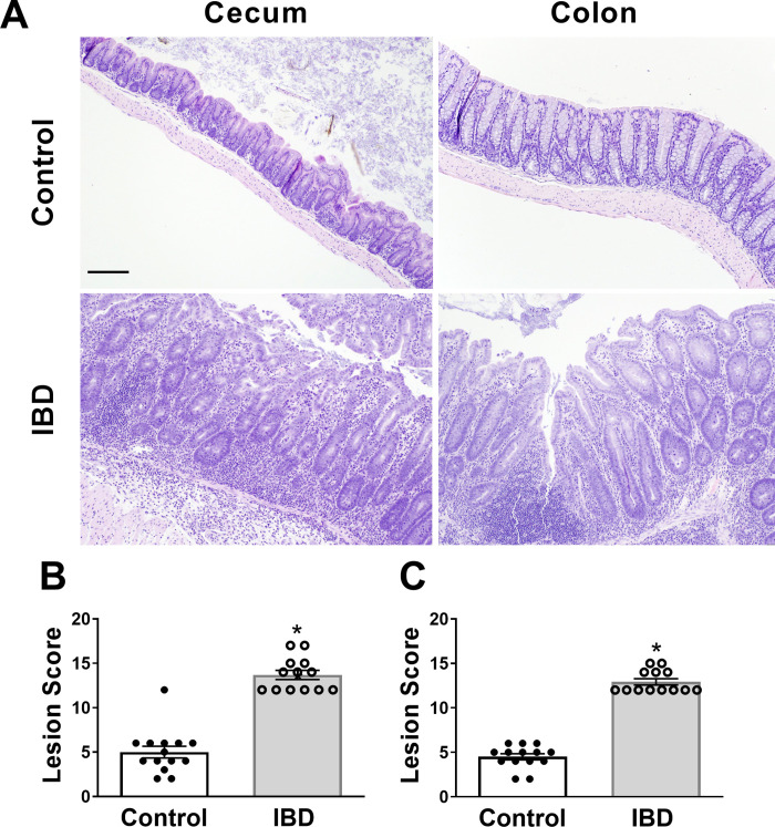
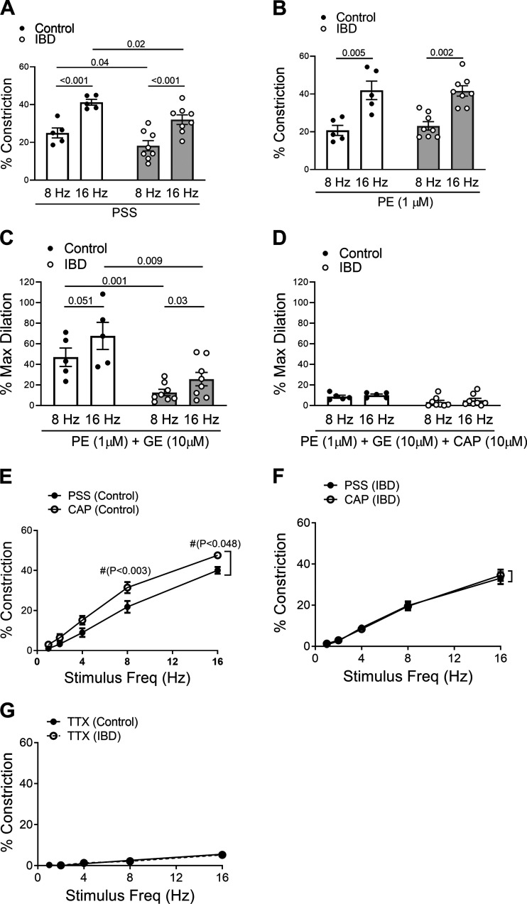
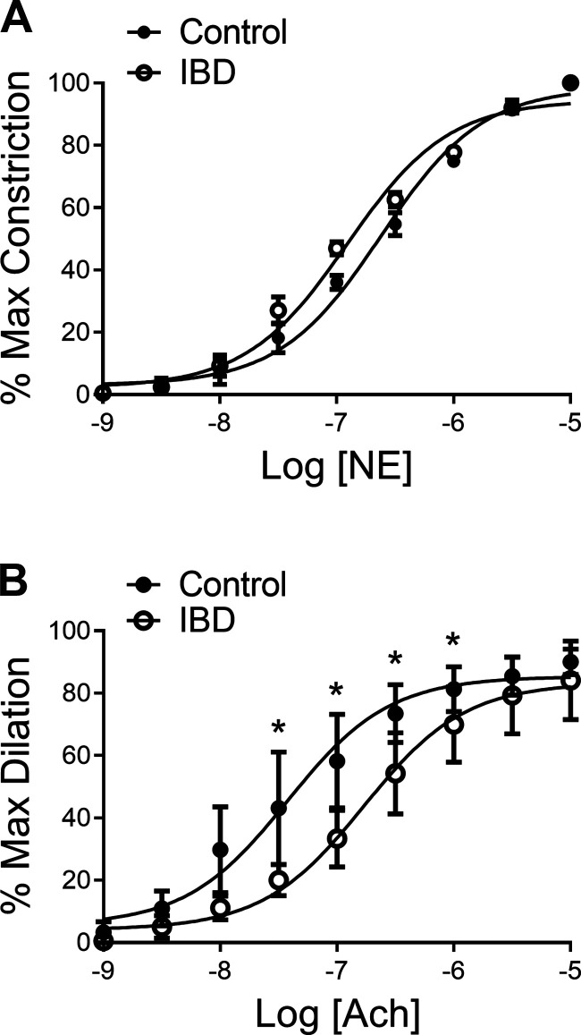
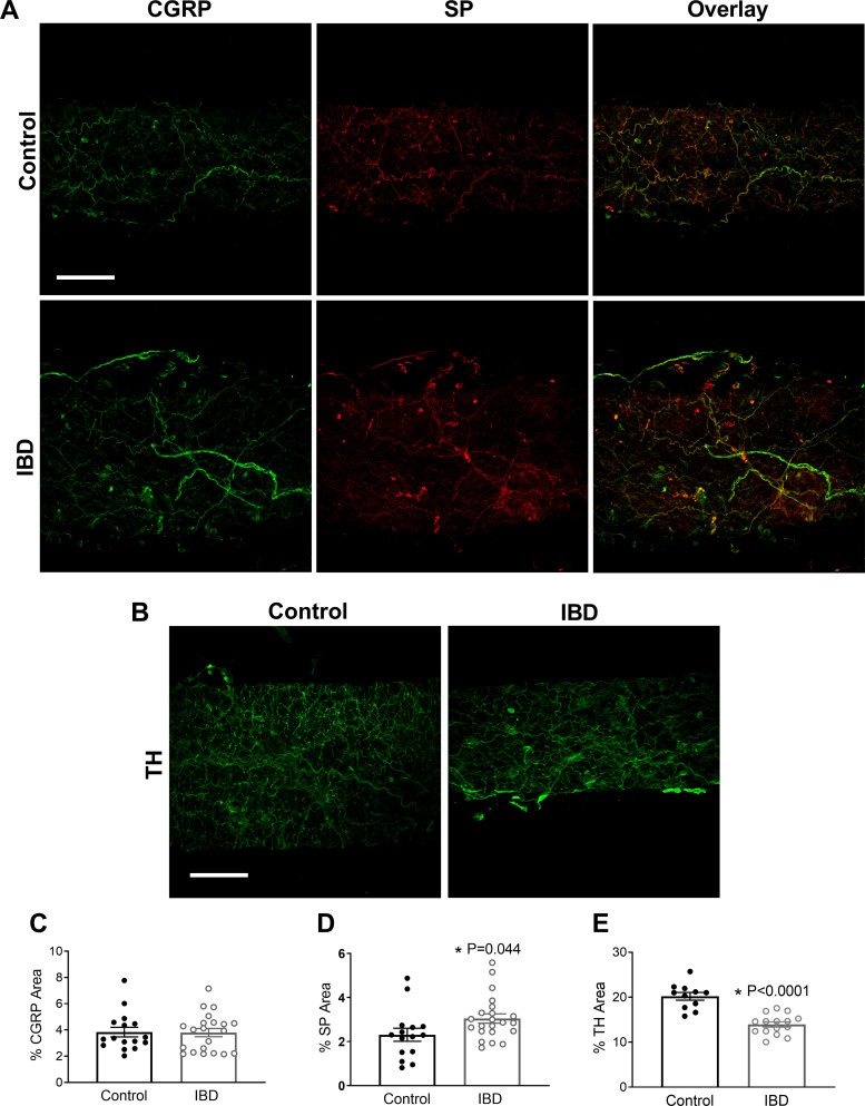
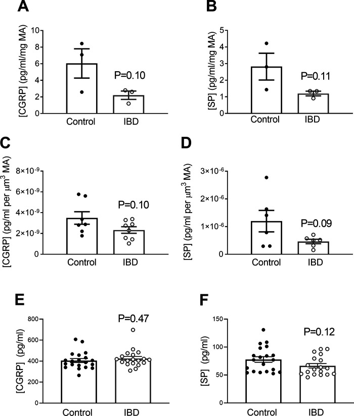
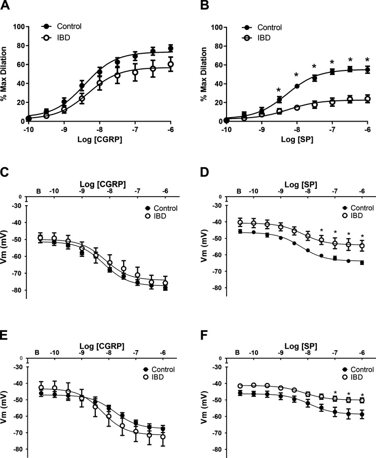
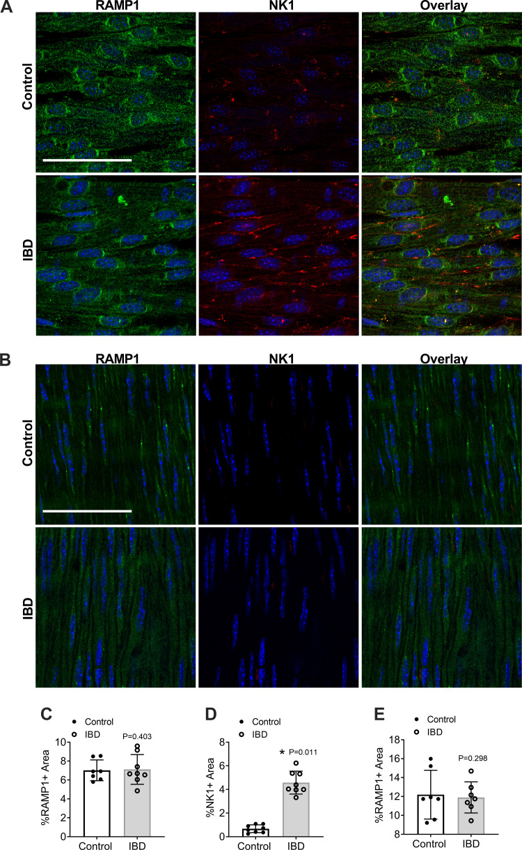
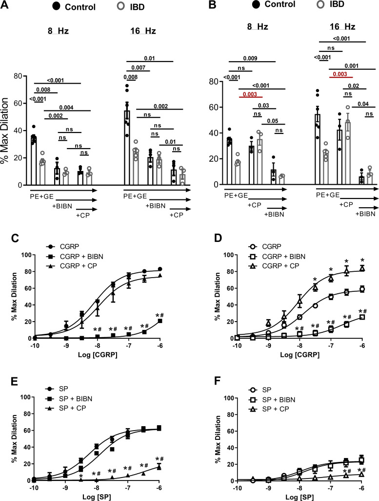
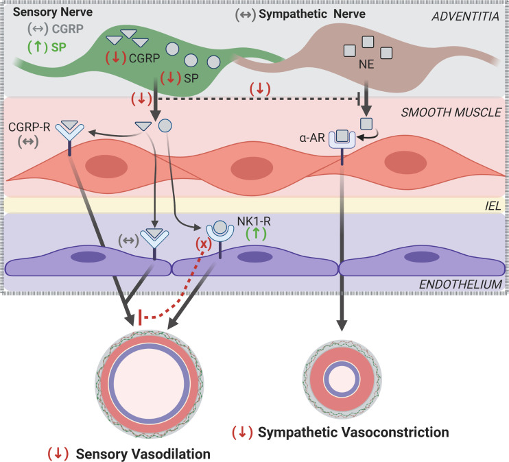
Inflammatory bowel disease (IBD) is associated with both impaired intestinal blood flow and increased risk of cardiovascular disease, but the functional role of perivascular nerves that control vasomotor function of mesenteric arteries (MAs) perfusing the intestine during IBD is unknown. Because perivascular sensory nerves and their transmitters calcitonin gene-related peptide (CGRP) and substance P (SP) are important mediators of both vasodilation and inflammatory responses, our objective was to identify IBD-related deficits in perivascular sensory nerve function and vascular neurotransmitter signaling. In MAs from an interleukin-10 knockout (IL-10-/-) mouse model, IBD significantly impairs electrical field stimulation (EFS)-mediated sensory vasodilation and inhibition of sympathetic vasoconstriction, despite decreased sympathetic nerve density and vasoconstriction. The MA content and EFS-mediated release of both CGRP and SP are decreased with IBD, but IBD has unique effects on each transmitter. CGRP nerve density, receptor expression, hyperpolarization, and vasodilation are preserved with IBD. In contrast, SP nerve density and receptor expression are increased, and SP hyperpolarization and vasodilation are impaired with IBD. A key finding is that blockade of SP receptors restores EFS-mediated sensory vasodilation and enhanced CGRP-mediated vasodilation in MAs from IBD but not Control mice. Together, these data suggest that an aberrant role for the perivascular sensory neurotransmitter SP and its downstream signaling in MAs underlies vascular dysfunction with IBD. We propose that with IBD, SP signaling impedes CGRP-mediated sensory vasodilation, contributing to impaired blood flow. Thus, substance P and NK1 receptors may represent an important target for treating vascular dysfunction in IBD.NEW & NOTEWORTHY Our study is the first to show that IBD causes profound impairment of sensory vasodilation and inhibition of sympathetic vasoconstriction in mesenteric arteries. This occurs alongside decreased SP-containing nerve density and increased expression of NK1 receptors for SP. In contrast, CGRP dilation, nerve density, and receptor expression are unchanged. Blocking NK1 receptors restores sensory vasodilation in MAs and increases CGRP-mediated vasodilation, indicating that SP interference with CGRP signaling may underlie impaired sensory vasodilation with IBD.

Keywords: adventitia; calcitonin gene-related peptide; inflammation; substance P; sympathetic nerves; vasodilation.


Conflict of interest statement

No conflicts of interest, financial or otherwise, are declared by the authors.

Figures

Figure 1.
Figure 1.
Helicobacter hepaticus-induced colitis in IL-10−/− mice. A: representative images of H&E-stained cecum and colon sections from Control (IL-10−/−) or IBD (H. hepaticus-inoculated IL-10−/−) mice. B and C: tissue lesion scores (means ± SE, range 0–24) in (B) cecum and (C) colon tissues. n = 14/group (Control: 8 males, 6 females; IBD: 7 males, 7 females). *P < 0.0001 vs. Control for both cecum and colon via unpaired t test. Scale bar = 50 μm and applies to all parts. H&E, hematoxylin-eosin; IBD, inflammatory bowel disease.
Figure 2.
Figure 2.
IBD impairs sympathetic constriction, sensory dilation, and sensory inhibition of sympathetic nerves. A and B: electrical field stimulation (EFS)-induced (8 and 16 Hz) sympathetic constrictions of isolated, cannulated, and pressurized arteries in PSS (A) and following preconstriction with phenylephrine (PE, 1 μM) (B). C: EFS-induced (8 and 16 Hz) sensory dilation of phenylephrine- (PE, 1 μM) and guanethidine (GE, 10 μM)-treated arteries (D) that is blocked by capsaicin (CAP; 10 μM). For AD, n = 5 or 8 mice/group (Control: 3 males, 2 females; IBD: 5 females, 3 males). E and F: sensory inhibition of sympathetic constriction in Control (E) and IBD arteries (F), shown as difference in EFS-induced (1–16Hz) constriction in the presence and absence of CAP (10 μM). G: tetrodotoxin (TTX) inhibition of EFS-induced (1–16 Hz) constriction. Data are means ± SD % constriction or % max dilation. For EG, n = 7 mice/group (Control: 4 males, 3 females; IBD: 3 males, 4 females). Data were analyzed by two-way ANOVA with Tukey’s post hoc test. IBD, inflammatory bowel disease; PSS, physiological salt solution.
Figure 3.
Figure 3.
IBD desensitizes arteries to acetylcholine but not norepinephrine. A: data are means ± SD for percent maximum dilation (obtained in 0 Ca2+ PSS + 10 μM sodium nitroprusside, SNP) of isolated, cannulated pressurized arteries preconstricted with phenylephrine (1 μM) and treated with cumulative concentrations of acetylcholine (ACh, 1 nM–10 μM). B: data are means ± SD for percent maximum constriction (normalized to 100%) of isolated, cannulated pressurized arteries treated with cumulative concentrations of norepinephrine (NE, 1 nM–10 μM). *P < 0.05 vs. Control (P values for 10−7.5, 10−7, 10−6.5, and 10−6 are 0.013, 0.0009, 0.0003, and 0.0096, respectively, via two-way ANOVA with Tukey’s post hoc test). n = 6 mice/group (Control and IBD: 3 males, 3 females). IBD, inflammatory bowel disease; PSS, physiological salt solution.
Figure 4.
Figure 4.
IBD does not alter structural or mechanical properties of mesenteric arteries. Inner diameter (μm, A), outer diameter (μm, B), wall thickness (C), cross-sectional area (D), wall:lumen (E), incremental distensibility (F), circumferential wall stress (G), and circumferential wall strain (H) are similar in Control and IBD arteries. Data are means ± SD for each variable with analysis via two-way ANOVA with Tukey’s post hoc test. n = 6 mice/group (Control and IBD: 4 males, 2 females). IBD, inflammatory bowel disease.
Figure 5.
Figure 5.
Perivascular nerve density is altered with IBD. A and B: representative maximum z-projections through the adventitia showing labeling for calcitonin gene-related peptide (CGRP, left), substance P (SP, middle), and CGRP-SP overlay (right) in Control (top, A) and IBD (bottom) arteries (B). Tyrosine hydroxylase (TH) in Control (left) and IBD (right) arteries. Scale bar = 100 μm. (CE): quantitation of nerve density for (C) CGRP, (D) SP, and (E) TH. Data are means ± SD for percent fluorescence within the vessel wall. *P < 0.05 vs. Control via unpaired t test. n = 11 or 21 vessel segments from 4 mice/group (Control: 2 males, 2 females; IBD: 3 males, 1 females). IBD, inflammatory bowel disease.
Figure 6.
Figure 6.
IBD decreases sensory neurotransmitter content and release from MAs. IBD was associated with trends toward decreased CGRP and SP content in intact MAs (n = 3 replicates from 6 mice/group (A and B); Control: 2 males, 4 females; IBD: 3 males, 3 females) and decreased EFS-mediated CGRP and SP release into tissue bath measured by ELISA (n = 6 mice/group (C and D); Control: 2 males, 4 females; IBD: 3 males, 3 females). E and F: serum CGRP was similar and serum SP was slightly decreased in Control vs. IBD mice. n = 19 or 20 mice/group (Control: 10 males, 10 females; IBD: 9 males, 10 females). All data were analyzed via unpaired t test. CGRP, calcitonin gene-related peptide; EFS, electrical field stimulation; IBD, inflammatory bowel disease; MAs, mesenteric arteries; SP, substance P.
Figure 7.
Figure 7.
IBD impairs dilation and hyperpolarization to exogenous SP but not CGRP. A and B: vasodilation and (CD) membrane hyperpolarization of isolated, cannulated, and pressurized arteries exposed to cumulative concentrations of CGRP (A and C) and SP (B and D). E and F: membrane hyperpolarization of intact EC tubes exposed to cumulative concentrations of CGRP (E) and SP (F). Data are means ± SD for % max dilation (n = 9 or 11 vessels from 4–6 mice/group) and Vm (n = 4 mice/group. *P < 0.05 vs. Control. B, baseline Vm. In (B), P = 0.038 for 10−8.5M SP, and P < 0.0001 for all other significant values. In (D), P values for 10−7.5, 10−7, 10−6.5, and 10−6 SP are 0.006, 0.008, 0.006, and 0.006, respectively. In (F), P values for 10−7, 10−6.5, and 10−6 SP are 0.020, 0.012, and 0.013, respectively. All data were analyzed via 2-way ANOVA with Tukey’s post hoc tests. CGRP, calcitonin gene-related peptide; EC, endothelial cell; IBD, inflammatory bowel disease; SP, substance P.
Figure 8.
Figure 8.
IBD alters NK1 but not RAMP1 expression and localization in mesenteric arteries. Images are representative maximum z-projections through the endothelial (A) and smooth muscle cell layers (B) of en face mesenteric arteries showing labeling for CGRP receptors (receptor activity modifying protein 1 (RAMP1, left), neurokinin 1 receptors (NK1, middle), and RAMP1-NK1 overlay (right) in Control (top) and IBD (bottom) arteries. All images include a DAPI nuclear stain. Scale bar = 50 μm. CE show the quantitation of staining of fluorescence for RAMP1 in endothelial cells (C), NK1 in endothelial cells (D), and RAMP1 in smooth muscle cells (E). Data were analyzed by Student’s t test. n = 8 vessel segments from 4 mice/group (Control: 2 males, 2 females; IBD: 2 males, 2 females). CGRP, calcitonin gene-related peptide; IBD, inflammatory bowel disease.
Figure 9.
Figure 9.
Respective roles of CGRP and SP receptor activation in EFS- and neuropeptide-induced vasodilation. A and B: EFS-induced (8 and 16 Hz) sensory dilation of PE- (1 μM) and GE (10 μM)-treated arteries in the presence of the CGRP receptor antagonist BIBN 4096 (1 μM) and BIBN + the NK1 antagonist CP99994 (1 μM) (A) or CP + BIBN. Note that NK1 inhibition restores sensory vasodilation with IBD (red bars) (B). Data are means ± SE % maximum dilations. n = 4 or 6/group (Control: 2 males, 2 females; IBD: 2 males, 4 females). CF: vasodilation of isolated, cannulated, and pressurized Control (C and E) and IBD arteries (D and F) exposed to cumulative concentrations of CGRP (C and D) and SP (E and F) in the absence and presence of BIBN or CP. *P < 0.05 vs. CGRP or SP alone; #P < 0.05 vs. CGRP + CP or SP + BIBN. n = 3 or 6 mice/group (Control: 1 male, 2 females; IBD: 3 male, 3 females). All concentration-response data were analyzed via two-way ANOVA with Tukey’s post hoc tests. CGRP, calcitonin gene-related peptide; EFS, electrical field stimulation; GE, guanethidine; IBD, inflammatory bowel disease; NK1, neurokinin 1; PE, phenylephrine; SP, substance P.
Figure 10.
Figure 10.
Summary of findings. Schematic depicts perivascular nerve modulation of mesenteric artery diameter in health and IBD. Upon stimulation of sensory nerves in healthy mesenteric arteries, CGRP and SP are coreleased. CGRP binds to CGRP receptors on smooth muscle and endothelial cells, and SP binds to NK1 receptors in endothelial cells. Released CGRP and SP also inhibit neurotransmitter release from sympathetic nerves. Each of these pathways contribute to vasodilation. Activation of sympathetic nerves causes release of NE, which binds to α-ARs on smooth muscle cells that facilitate vasoconstriction. Items in parentheses indicate IBD-related changes indicated by the present study. Note that this schematic is limited in scope and only depicts nerves, transmitters, and receptors relevant to the present study. α-AR, alpha adrenergic receptor; CGRP-R, calcitonin gene-related peptide receptor; IBD, inflammatory bowel disease; IEL, internal elastic lamina; NE, norepinephrine; NK1-R, neurokinin 1 receptor; SP, substance P. Figure was created using BioRender.

Similar articles

Cited by

References

    1. Papa A, Danese S, Urgesi R, Grillo A, Guglielmo S, Roberto I, Bonizzi M, Guidi L, De Vitis I, Santoliquido A, Fedeli G, Gasbarrini G, Gasbarrini A. Early atherosclerosis in patients with inflammatory bowel disease. Eur Rev Med Pharmacol Sci 10: 7–11, 2006. - PubMed
    1. Zanoli L, Cannavò M, Rastelli S, Di Pino L, Monte I, Di Gangi M, Boutouyrie P, Inserra G, Laurent S, Castellino P. Arterial stiffness is increased in patients with inflammatory bowel disease. J Hypertens 30: 1775–1781, 2012. doi:10.1097/HJH.0b013e3283568abd. - DOI - PubMed
    1. Miehsler W, Reinisch W, Valic E, Osterode W, Tillinger W, Feichtenschlager T, Grisar J, Machold K, Scholz S, Vogelsang H, Novacek G. Is inflammatory bowel disease an independent and disease specific risk factor for thromboembolism? Gut 53: 542–548, 2004. doi:10.1136/gut.2003.025411. - DOI - PMC - PubMed
    1. Kristensen SL, Ahlehoff O, Lindhardsen J, Erichsen R, Lamberts M, Khalid U, Nielsen OH, Torp-Pedersen C, Gislason GH, Hansen PR. Inflammatory bowel disease is associated with an increased risk of hospitalization for heart failure: a Danish nationwide cohort study. Circ Heart Fail 7: 717–722, 2014. doi:10.1161/CIRCHEARTFAILURE.114.001152. - DOI - PubMed
    1. Aniwan S, Pardi DS, Tremaine WJ, Loftus EV. Jr, Increased risk of acute myocardial infarction and heart failure in patients with inflammatory bowel diseases. Clin Gastroenterol Hepatol 16: 1607–1615, 2018. doi:10.1016/j.cgh.2018.04.031. - DOI - PMC - PubMed

Publication types

MeSH terms

LinkOut - more resources