. 2021 Mar 12;13(2):025004.
doi: 10.1088/1758-5090/abce0a.

Multiplexing physical stimulation on single human induced pluripotent stem cell-derived cardiomyocytes for phenotype modulation

Affiliations

Multiplexing physical stimulation on single human induced pluripotent stem cell-derived cardiomyocytes for phenotype modulation

Worrapong Kit-Anan et al. Biofabrication. .

Abstract

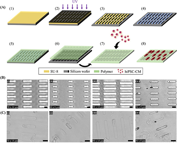
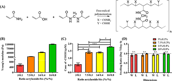
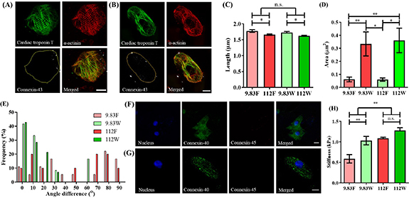
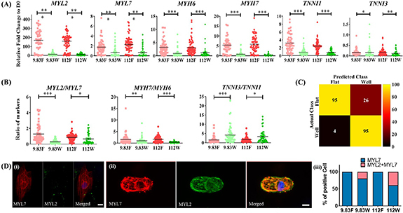
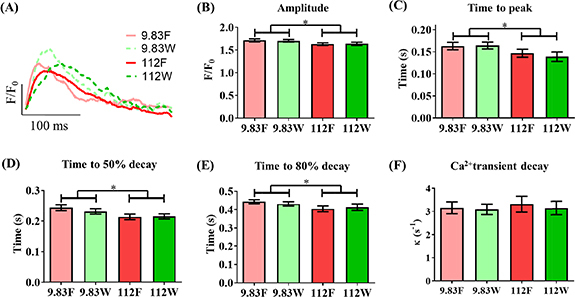
Traditional in vitro bioengineering approaches whereby only individual biophysical cues are manipulated at any one time are highly inefficient, falling short when recapitulating the complexity of the cardiac environment. Multiple biophysical cues are present in the native myocardial niche and are essential during development, as well as in maintenance of adult cardiomyocyte (CM) phenotype in both health and disease. This study establishes a novel biofabrication workflow to study and manipulate hiPSC-CMs and to understand how these cells respond to a multiplexed biophysical environment, namely 3D shape and substrate stiffness, at a single cell level. Silicon masters were fabricated and developed to generate inverse patterns of the desired 3D shapes in bas relief, which then were used to mold the designed microwell arrays into a hydrogel. Polyacrylamide (PAAm) was modified with the incorporation of acrylic acid to provide a carboxylic group conjugation site for adhesion motifs, without compromising capacity to modulate stiffness. In this manner, two individual parameters can be finely tuned independently within the hydrogel: the shape of the 3D microwell and its stiffness. The design allows the platform to isolate single hiPSC-CMs to study solely biophysical cues in the absence of cell-cell physical interaction. Under physiologic-like physical conditions (3D shape resembling that of adult CM and 9.83 kPa substrate stiffness that mimics muscle stiffness), isolated single hiPSC-CMs exhibit increased Cx-43 density, cell membrane stiffness and calcium transient amplitude; co-expression of the subpopulation-related MYL2-MYL7 proteins; and higher anisotropism than cells in pathologic-like conditions (flat surface and 112 kPa substrate stiffness). This demonstrates that supplying a physiologic or pathologic microenvironment to an isolated single hiPSC-CM in the absence of any physical cell-to-cell communication in this biofabricated platform leads to a significantly different set of cellular features, thus presenting a differential phenotype. Importantly, this demonstrates the high plasticity of hiPSC-CMs even in isolation. The ability of multiple biophysical cues to significantly influence isolated single hiPSC-CM phenotype and functionality highlights the importance of fine-tuning such cues for specific applications. This has the potential to produce more fit-for-purpose hiPSC-CMs. Further understanding of human cardiac development is enabled by the robust, versatile and reproducible biofabrication techniques applied here. We envision that this system could be easily applied to other tissues and cell types where the influence of cellular shape and stiffness of the surrounding environment is hypothesized to play an important role in physiology.


Figures

Figure 1.
Figure 1.
Microfabrication of polyacrylamide-co-acrylic acid (PAAm-co-PAAc) hydrogel and cell seeding. (A) Schematic representation of the microfabrication and cell-seeding workflow [1]: spin coating of photoresist (SU-8) [2], pattern transfer from photomask to photoresist by UV [3], pattern resulting from photolithography: arrays of pillars resembling the shape of adult CM [4], perfluorosilanization of substrate [5], PAAm-co-PAAc coating of the hard mold by drop-casting [6], removal of PAAm-co-PAAc and ECM conjugation [7], hiPSC-CMs seeding on the platform [8], hiPSC-CM culture on micropatterned PAAm-co-PAAc hydrogel. (B) Representative brightfield images of PAAm-co-PAAc hydrogels of (i) 70 × 14 µm, (ii) 40 × 8 µm, (iii) 98 × 14 µm and (iv) 56 × 8 µm. (C) Representative brightfield images of hiPSC-CMs cultured on (i) 70 × 14 µm, (ii) 40 × 8 µm, (iii) 98 × 14 µm and (iv) 56 × 8 µm. Scale bars: 25 µm.
Figure 2.
Figure 2.
PAAm-co-PAAc hydrogel characterizations. (A) Chemical structure of PAAm-co-PAAc. (B) Young’s modulus measurement by contact-mode AFM. (C) Available carboxylic groups measured using Toluidine Blue O. (ANOVA, *p < 0.05, ***p < 0.001). (D) Swelling ratio of the 3D microwells showing no significant effect on the width (W) and length (L) directions after 7 d incubation in cell culture media (Student T-test, no significant differences). Dashed line represents no change (Swelling ratio = 1). Data shown as mean ± SEM. N = 3, independent experiments.
Figure 3.
Figure 3.
Concomitant modulation of cell shape and stiffness by PAAm-co-PAAc hydrogel influences the structure of isolated single hiPSC-CMs. Representative images of hiPSC-CMs stained for cTnT, α-actinin and Cx-43 on (A) the flat control surface and (B) inside the 3D adult-like microwell of physiologic substrate stiffness (9.83 kPa). Yellow outline demonstrates the perimeter of cell. (C) Sarcomere length showed a significantly higher spacing when cultured on physiologic stiffness than on pathologic surfaces (112 kPa) (2-way ANOVA, *p < 0.05). (D) Quantification of area occupied by Cx-43 on 3D microwells and flat surface controls, showing increased presence of Cx-43 in cells residing inside 3D microwells (2-way ANOVA, *p< 0.05, **p < 0.01). (E) Analysis of CM sarcomeric directionality, measured against X-axis, demonstrates capacity of the 3D microwells for inducing cell alignment. Representative images of Cx-40 and Cx-45 staining on (F) the flat control surface and (G) 3D microwell of physiologic stiffness. (H) Cell membrane stiffness measured using AFM, demonstrating increased stiffness under pathologic condition versus physiologic condition, as well as increased membrane rigidity of isolated single hiPSC-CMs residing inside the 3D microwells (2-way ANOVA, **p < 0.01). Scale bars: 10 µm. Data shown as mean ± SEM, N ⩾ 3. (9.83 F = physiologic stiffness flat control, 9.83 W = physiologic stiffness 3D microwell, 112 F = pathologic stiffness flat control, 112 W = pathologic stiffness 3D microwell).
Figure 4.
Figure 4.
Multiplexing cell shape and substrate stiffness modulates gene and protein expression in isolated single hiPSC-CMs. (A) Single cell RT-qPCR analysis of the expression levels of cardiac structural markers, showing upregulated levels of MYL2, MYL7, MYH6, MYH7, TNNI1 and downregulated expression of TNNI3 in control flat surfaces with different substrate stiffnesses (2-way ANOVA, *p < 0.05, **p < 0.01, ***p < 0.001). (B) Ratio of cardiac structural markers. Values were normalized to hiPSC-CMs day 0 before plating. (2-way ANOVA, *p < 0.05, **p < 0.01, ***p < 0.001). Data shown as mean ± SEM. N = 4, n ⩾ 42. (C) Confusion matrix heatmap using a generalized linear model (GLM) of predicted class versus actual class of individual single hiPSC-CMs in each condition (Influences of biophysical cues provide on gene expression). (D) Immunostaining of hiPSC-CMs for MYL2 and MYL7, cultured on (a) flat controls, (b) 3D microwells on the platform and (c) bar graph showing the proportion of MYL2 and co-expression of MYL2 and MYL7 cultured on different conditions. Scale bars: 10 µm. (9.83 F = physiologic stiffness flat control, 9.83 W = physiologic stiffness 3D microwell, 112 F = pathologic stiffness flat control, 112 W = pathologic stiffness 3D microwell).
Figure 5.
Figure 5.
Substrate stiffness dominates influence on Ca2+ handling of isolated single hiPSC-CMs over cell shape. Ca2+ handling was assessed by using an isogenic hiPSC line harboring a genetically encoded calcium indicator, GCaMP6f, and optical mapping under field stimulation at 1 Hz. (A) Representative trace of intracellular Ca2+. Ca2+ transient parameters: (B) amplitude, (C) time to peak, (D) time to 50% decay, (E) time to 80% decay and (F) Ca2+ Transient decay. Data shown as mean ± SEM. N = 4 independent experiments, n ⩾ 36 (2-way ANOVA, *p < 0.05). 9.83 F = physiologic stiffness flat control, 9.83 W = physiologic stiffness 3D microwell, 112 F = pathologic stiffness flat control, 112 W = pathologic stiffness 3D microwell.

References

    1. Mozaffarian D, et al. Heart disease and stroke statistics—2016 update. Circulation. 2016;133:1–324. doi: 10.1161/CIR.0000000000000350. - DOI - PubMed
    1. Kikuchi K, Poss K D. Cardiac regenerative capacity and mechanisms. Annu. Rev. Cell Dev. Biol. 2012;28:719–41. doi: 10.1146/annurev-cellbio-101011-155739. - DOI - PMC - PubMed
    1. Takahashi K, Tanabe K, Ohnuki M, Narita M, Ichisaka T, Tomoda K, Yamanaka S. Induction of pluripotent stem cells from adult human fibroblasts by defined factors. Cell. 2007;131:861–72. doi: 10.1016/j.cell.2007.11.019. - DOI - PubMed
    1. Lian X, Zhang J, Azarin S M, Zhu K, Hazeltine L B, Bao X, Hsiao C, Kamp T J, Palecek S P. Directed cardiomyocyte differentiation from human pluripotent stem cells by modulating Wnt/β-catenin signaling under fully defined conditions. Nat. Protocols. 2013;8:162–75. doi: 10.1038/nprot.2012.150. - DOI - PMC - PubMed
    1. Wang B, Kit-Anan W, Terracciano C. Many cells make life work—multicellularity in stem cell-based cardiac disease modelling. Int. J. Mol. Sci. 2018;19:3361. doi: 10.3390/ijms19113361. - DOI - PMC - PubMed

Publication types

LinkOut - more resources