

Sialadenoma papilliferum: clinicopathologic, Immunohistochemical, molecular analyses of new five cases and review of the literature
- PMID: 33712056
- PMCID: PMC7953575
- DOI: 10.1186/s13000-021-01084-5
Sialadenoma papilliferum: clinicopathologic, Immunohistochemical, molecular analyses of new five cases and review of the literature
Abstract
Background: Sialadenoma papilliferum (SP) is an extremely rare benign neoplasm of salivary glands. To explore and define the clinicopathological features of SP, we retrospectively analyzed 89 cases previously reported and five new cases.
Methods: The clinical features, histopathology, immunohistochemistry and molecular analysis of our cases were further performed and the related literatures were reviewed and analyzed.
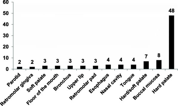
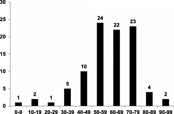
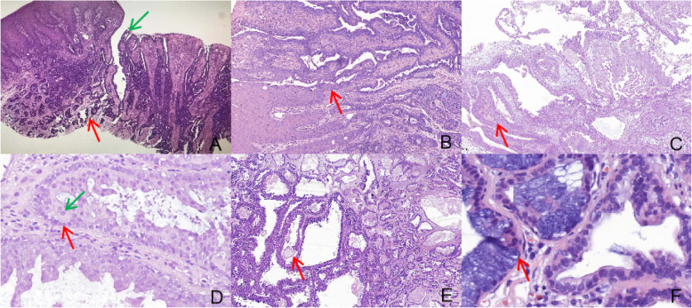
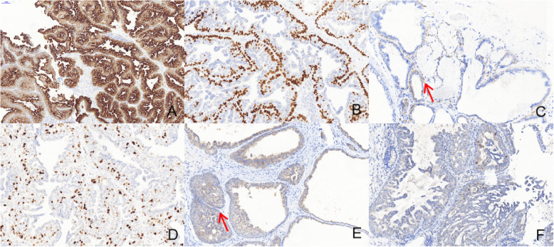
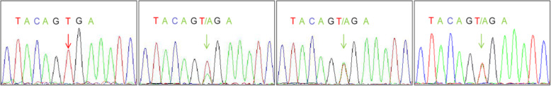
Results: Combining 89 cases from the literature with our cases, the hard palate was the most common locations for SP. However, two of our cases were rarely located in the esophageal mucosa. Among all cases, the male gender was more affected, with the average age and median age of 61.8 and 62 years, respectively. Conventional histomorphologically, SP was characterized by complex papillary structures with a biphasic growth pattern of exophytic squamous component and endophytic glandular component. The glandular structures were lined by a double layer of epithelium composed of flattened or cuboidal basal cells and a cuboidal or columnar luminal cells formed papillary infoldings into the ductal lumina. Immunohistochemically, the luminal epithelial configurations showed strong expression of CK7 along the luminal cell membrane, while the basal myoepithelia displayed strong nuclear p63 expression. In both the glandular and squamous tumour components showed BRAF V600E-positive immunostaining and BRAF V600E mutation.
Conclusion: For the first time, we have comprehensively aggregated and analyzed 90 cases sialadenoma papilliferum from almost all previous publications, and further explored the clinicopathological features of SP; concordantly, this study demonstrated that SP shows a papillomatous growth pattern with exophytic and endophytic proliferation of ductal epithelium composed of double-layered cells harboring BRAF V600E mutation. Additionly, adequate treatment for SP is surgical excision, with a favorable prognosis in patients.
Keywords: BRAF; Clinical features; Histopathology; Immunohistochemistry; Sialadenoma papilliferum.
Conflict of interest statement
The authors declared no potential conflicts of interest with respect to the research, authorship, and/or publication of this article.
Figures
Similar articles
-
Salivary Sialadenoma Papilliferum Consists of Two Morphologically, Immunophenotypically, and Genetically Distinct Subtypes.Head Neck Pathol. 2020 Jun;14(2):489-496. doi: 10.1007/s12105-019-01068-4. Epub 2019 Aug 31. Head Neck Pathol. 2020. PMID: 31473937 Free PMC article.
-
Clinicopathologic Study of Sialadenoma Papilliferum of the Minor Salivary Glands: A Series of 8 New Cases With BRAF V600E Mutation-specific Immunohistochemical Analysis.Int J Surg Pathol. 2023 Oct;31(7):1265-1272. doi: 10.1177/10668969221147170. Epub 2023 Jan 11. Int J Surg Pathol. 2023. PMID: 36632022
-
Sialadenoma Papilliferum.Surg Pathol Clin. 2021 Mar;14(1):43-51. doi: 10.1016/j.path.2020.09.006. Epub 2021 Jan 5. Surg Pathol Clin. 2021. PMID: 33526222 Review.
-
Histopathological evaluation of minor salivary gland papillary-cystic tumours: focus on genetic alterations in sialadenoma papilliferum and intraductal papillary mucinous neoplasm.Histopathology. 2020 Feb;76(3):411-422. doi: 10.1111/his.13990. Epub 2019 Dec 1. Histopathology. 2020. PMID: 31505033
-
Ductal papillomas of salivary gland origin: A report of 19 cases and a review of the literature.Oral Surg Oral Med Oral Pathol Oral Radiol Endod. 2001 Jul;92(1):68-77. doi: 10.1067/moe.2001.115978. Oral Surg Oral Med Oral Pathol Oral Radiol Endod. 2001. PMID: 11458248 Review.
Cited by
-
Salivary Duct Carcinoma with Rhabdoid Features of the Parotid Gland with No E-Cadherin Expression: A Report with Anti-HER2 Therapy and Review of the Literature.Dent J (Basel). 2023 Sep 25;11(10):229. doi: 10.3390/dj11100229. Dent J (Basel). 2023. PMID: 37886914 Free PMC article.
-
Bronchial salivary gland-type intraductal carcinoma with KIAA1217::RET gene fusion composed of intercalated and oncocytic components.Virchows Arch. 2023 Apr;482(4):789-795. doi: 10.1007/s00428-022-03456-8. Epub 2022 Nov 21. Virchows Arch. 2023. PMID: 36414804
-
Low-grade non-intestinal-type sinonasal adenocarcinoma: a histologically distinctive but molecularly heterogeneous entity.Mod Pathol. 2022 Sep;35(9):1160-1167. doi: 10.1038/s41379-022-01068-w. Epub 2022 Mar 23. Mod Pathol. 2022. PMID: 35322195
-
Sialadenoma papilliferum of a minor salivary gland with transformation to mucoepidermoid carcinoma.J Postgrad Med. 2022 Apr-Jun;68(2):112-114. doi: 10.4103/jpgm.jpgm_912_21. J Postgrad Med. 2022. PMID: 35381752 Free PMC article.
-
Sialadenoma Papilliferum: An Uncommon Verruco-Papillary Lesion of the Oral Cavity.Indian J Otolaryngol Head Neck Surg. 2025 Feb;77(2):999-1002. doi: 10.1007/s12070-024-05236-z. Epub 2024 Nov 27. Indian J Otolaryngol Head Neck Surg. 2025. PMID: 40065975
References
MeSH terms
Grants and funding
LinkOut - more resources
Full Text Sources
Other Literature Sources
Medical
Research Materials
