

Value of conventional magnetic resonance imaging texture analysis in the differential diagnosis of benign and borderline/malignant phyllodes tumors of the breast
- PMID: 33712070
- PMCID: PMC7953576
- DOI: 10.1186/s40644-021-00398-3
Value of conventional magnetic resonance imaging texture analysis in the differential diagnosis of benign and borderline/malignant phyllodes tumors of the breast
Abstract
Background: The purpose of this study was to determine the potential value of magnetic resonance imaging (MRI) texture analysis (TA) in differentiating between benign and borderline/malignant phyllodes tumors of the breast.
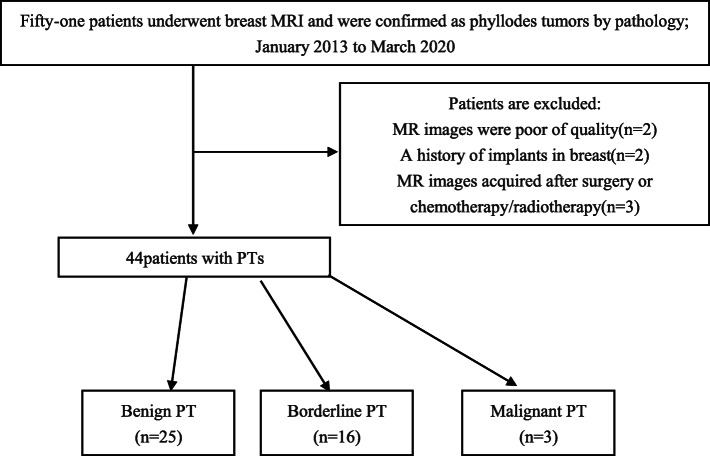
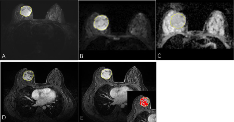
Methods: The preoperative MRI data of 25 patients with benign phyllodes tumors (BPTs) and 19 patients with borderline/malignant phyllodes tumors (BMPTs) were retrospectively analyzed. A gray-level histogram and gray-level cooccurrence matrix (GLCM) were used for TA with fat-suppressed T2-weighted imaging (FS-T2WI), diffusion-weighted imaging (DWI), apparent diffusion coefficient (ADC) images, and 2- and 7-min postcontrast T1W images on dynamic contrast-enhanced MRI (DCE-T1WI2min and DCE-T1WI7min) between BPTs and BMPTs. Independent sample t-test and Mann-Whitney U test were performed for intergroup comparison. A regression model was established by using binary logistic regression analysis, and receiver operating characteristic (ROC) curve analysis was carried out to evaluate diagnostic efficiency.
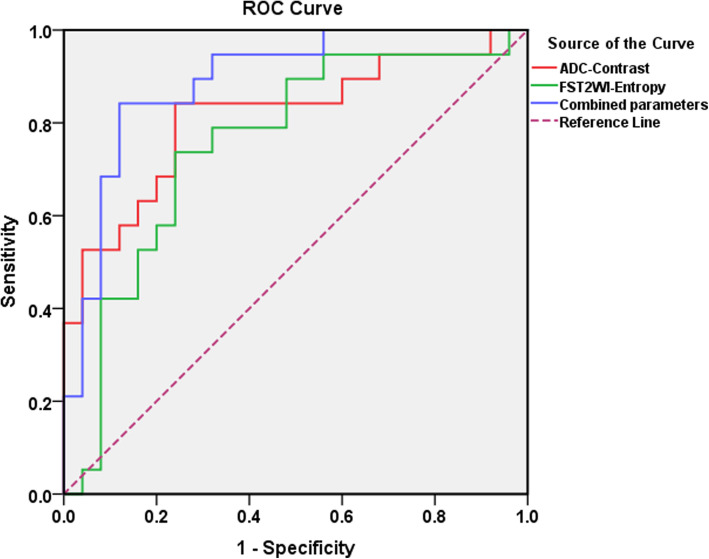
Results: For ADC images, the texture parameters angular second moment (ASM), correlation, contrast, entropy and the minimum gray values of ADC images (ADCMinimum) showed significant differences between the BPT group and BMPT group (all p<0.05). The parameter entropy of FS-T2WI and the maximum gray values and kurtosis of the tumor solid region of DCE-T1WI7min also showed significant differences between these two groups. Except for ADCMinimum, angular second moment of FS-T2WI (FS-T2WIASM), and the maximum gray values of DCE-T1WI7min (DCE-T1WI7min-Maximum) of the tumor solid region, the AUC values of other positive texture parameters mentioned above were greater than 0.75. Binary logistic regression analysis demonstrated that the contrast of ADC images (ADCContrast) and entropy of FS-T2WI (FS-T2WIEntropy) could be considered independent texture variables for the differential diagnosis of BPTs and BMPTs. Combined, the AUC of these parameters was 0.891 (95% CI: 0.793-0.988), with a sensitivity of 84.2% and a specificity of up to 89.0%.
Conclusion: Texture analysis could be helpful in improving the diagnostic efficacy of conventional MR images in differentiating BPTs and BMPTs.
Keywords: Differential diagnosis; Magnetic resonance imaging; Phyllodes tumors; Texture analysis.
Conflict of interest statement
The authors declare that they have no competing interest.
Figures
Similar articles
-
The Potential Value of Texture Analysis Based on Dynamic Contrast-Enhanced MR Images in the Grading of Breast Phyllode Tumors.Front Oncol. 2021 Nov 10;11:745242. doi: 10.3389/fonc.2021.745242. eCollection 2021. Front Oncol. 2021. PMID: 34858821 Free PMC article.
-
Pretreatment Multiparametric MRI-Based Radiomics Analysis for the Diagnosis of Breast Phyllodes Tumors.J Magn Reson Imaging. 2023 Feb;57(2):633-645. doi: 10.1002/jmri.28286. Epub 2022 Jun 3. J Magn Reson Imaging. 2023. PMID: 35657093
-
Can whole-tumor apparent diffusion coefficient histogram analysis be helpful to evaluate breast phyllode tumor grades?Eur J Radiol. 2019 May;114:25-31. doi: 10.1016/j.ejrad.2019.02.035. Epub 2019 Feb 27. Eur J Radiol. 2019. PMID: 31005172
-
MRI in differentiation of benign and malignant tongue tumors.Front Biosci (Landmark Ed). 2015 Jan 1;20(4):614-20. doi: 10.2741/4326. Front Biosci (Landmark Ed). 2015. PMID: 25553468 Review.
-
Ovarian solid tumors: MR imaging features with radiologic-pathologic correlation.Jpn J Radiol. 2020 Aug;38(8):719-730. doi: 10.1007/s11604-020-00976-8. Epub 2020 Apr 27. Jpn J Radiol. 2020. PMID: 32342277 Review.
Cited by
-
MRI-based radiomics analysis for differentiating phyllodes tumors of the breast from fibroadenomas.Eur Radiol. 2022 Jun;32(6):4090-4100. doi: 10.1007/s00330-021-08510-8. Epub 2022 Jan 19. Eur Radiol. 2022. PMID: 35044510
-
Third International Consensus Conference on lesions of uncertain malignant potential in the breast (B3 lesions).Virchows Arch. 2023 Jul;483(1):5-20. doi: 10.1007/s00428-023-03566-x. Epub 2023 Jun 17. Virchows Arch. 2023. PMID: 37330436 Free PMC article. Review.
-
The Potential Value of Texture Analysis Based on Dynamic Contrast-Enhanced MR Images in the Grading of Breast Phyllode Tumors.Front Oncol. 2021 Nov 10;11:745242. doi: 10.3389/fonc.2021.745242. eCollection 2021. Front Oncol. 2021. PMID: 34858821 Free PMC article.
-
Navigating the Uncertainty of B3 Breast Lesions: Diagnostic Challenges and Evolving Management Strategies.J Pers Med. 2025 Jan 18;15(1):36. doi: 10.3390/jpm15010036. J Pers Med. 2025. PMID: 39852228 Free PMC article. Review.
-
Differentiation between Phyllodes Tumors and Fibroadenomas through Breast Ultrasound: Deep-Learning Model Outperforms Ultrasound Physicians.Sensors (Basel). 2023 May 26;23(11):5099. doi: 10.3390/s23115099. Sensors (Basel). 2023. PMID: 37299826 Free PMC article.
References
-
- Bendifallah S, Canlorbe G. [common benign breast tumors including fibroadenoma, phyllodes tumors, and papillary lesions: guidelines]. J Gynecol Obstet. Biol Reprod (Paris) 2015;44(10):1017–1029. - PubMed
MeSH terms
LinkOut - more resources
Full Text Sources
Other Literature Sources
Medical
