. 2021 Mar 12;9(1):41.
doi: 10.1186/s40478-021-01141-6.

Tau assemblies do not behave like independently acting prion-like particles in mouse neural tissue

Affiliations

Tau assemblies do not behave like independently acting prion-like particles in mouse neural tissue

Lauren V C Miller et al. Acta Neuropathol Commun. .

Abstract

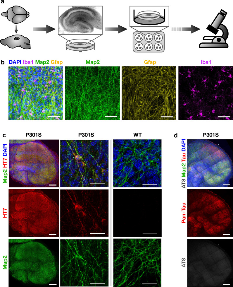
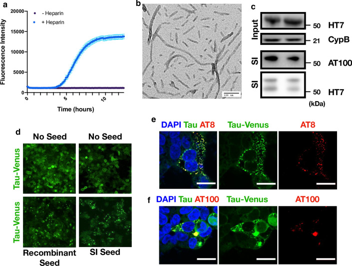
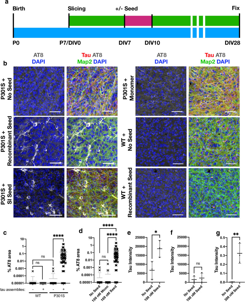
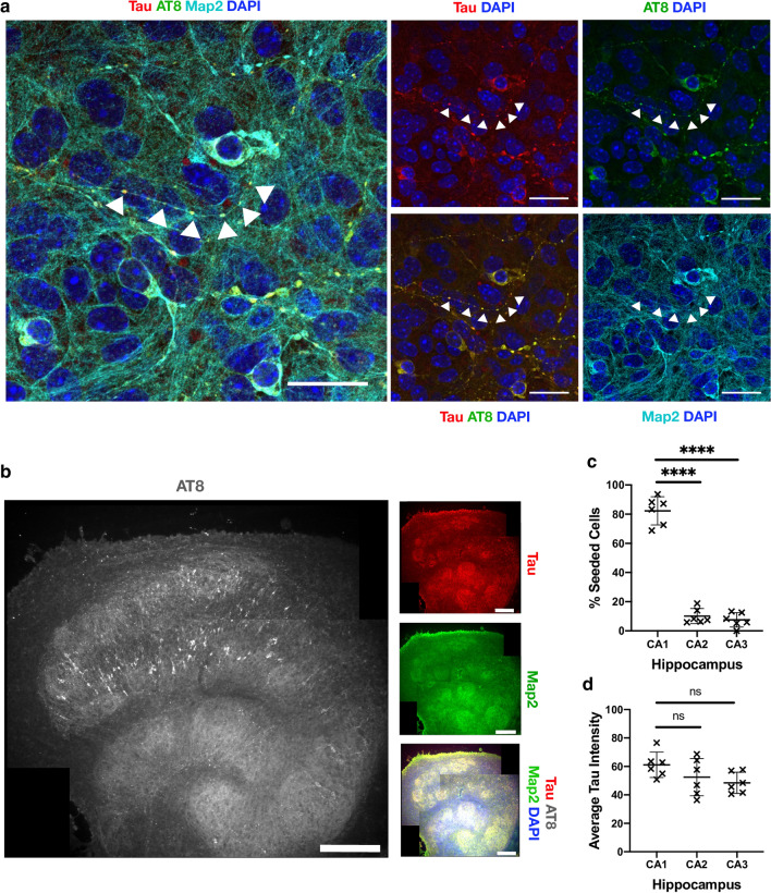
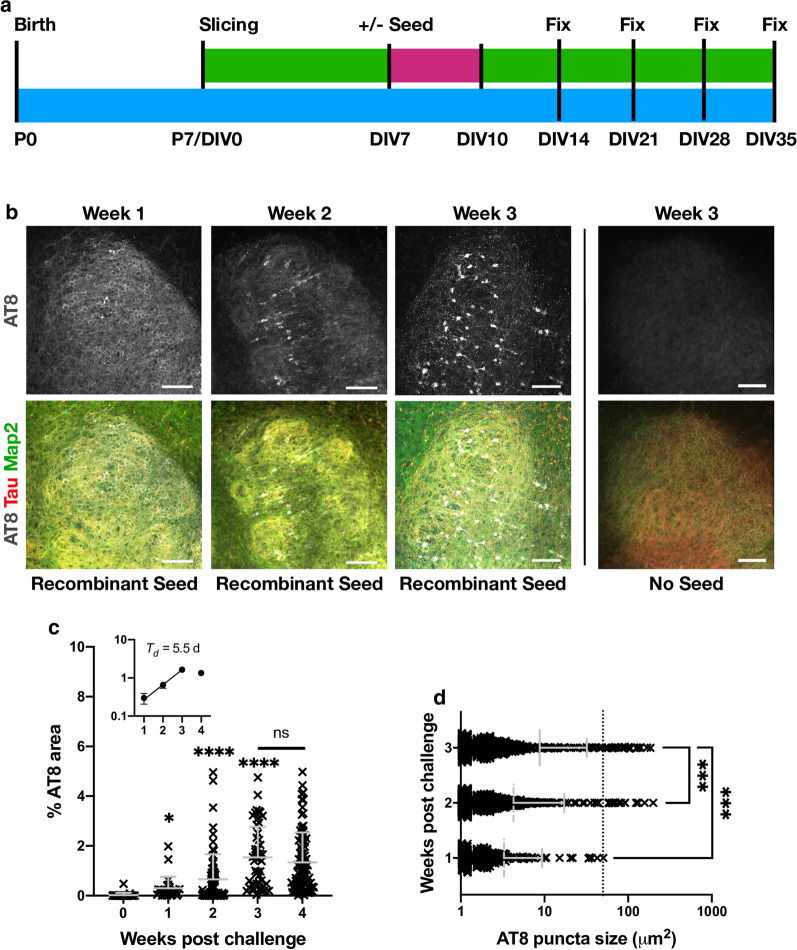
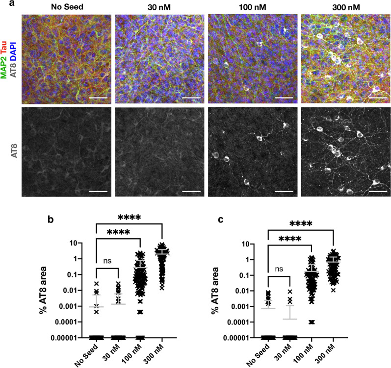
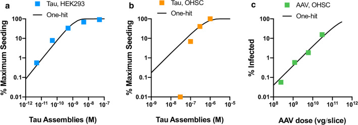
A fundamental property of infectious agents is their particulate nature: infectivity arises from independently-acting particles rather than as a result of collective action. Assemblies of the protein tau can exhibit seeding behaviour, potentially underlying the apparent spread of tau aggregation in many neurodegenerative diseases. Here we ask whether tau assemblies share with classical pathogens the characteristic of particulate behaviour. We used organotypic hippocampal slice cultures from P301S tau transgenic mice in order to precisely control the concentration of extracellular tau assemblies in neural tissue. Whilst untreated slices displayed no overt signs of pathology, exposure to recombinant tau assemblies could result in the formation of intraneuronal, hyperphosphorylated tau structures. However, seeding ability of tau assemblies did not titrate in a one-hit manner in neural tissue. The results suggest that seeding behaviour of tau arises at high concentrations, with implications for the interpretation of high-dose intracranial challenge experiments and the possible contribution of seeded aggregation to human disease.

Keywords: Neurodegeneration; Organotypic hippocampal slice cultures; Prion-like activity; Tau seeded aggregation; Tauopathies.


Conflict of interest statement

The authors declare that they have no competing interests.

Figures

Fig. 1
Fig. 1
Organotypic hippocampal slice cultures (OHSCs) maintain cellular diversity and display no spontaneous tau pathology. a Schematic of the production of OHSCs. 300 µm thick sagittal slices are cut from brain hemispheres. The hippocampus is then dissected and cultured at the air–liquid interface on semi-permeable membranes with media supplied to the basal side of the membrane. b OHSCs from mice transgenic for P301S tau were fixed after 2 weeks in culture and stained for nuclei (DAPI), the neuronal marker Map2, the astrocyte marker Gfap, and the microglial marker Iba1. Scale bars, 50 µm. c OHSCs from P301S tau transgenic mice stain positive for human tau-specific antibody HT7 whereas OHSCs from WT mice do not. Scale bars, 250 µm and 25 µm. d OHSCs from P301S tau transgenic mice after 5 weeks in culture display only background levels of staining with the phospho-tau specific antibody AT8. Slices were stained with DAPI and Map2 as above and with pan-tau. Scale bars, 250 µm
Fig. 2
Fig. 2
Characterisation of seed competent tau assemblies. a Time course of recombinant P301S tau assembly, monitored by Thioflavin T fluorescence. b Representative transmission electron microscopy image of heparin assembled P301S tau. c Aged P301S tau transgenic mouse brain homogenate was immunoblotted for human tau (HT7 antibody) to detect transgenic tau and for cyclophilin B which served as a loading control. Presence of sarkosyl insoluble (SI) tau was confirmed with the HT7 antibody and AT100 (tau phosphorylated at pT212, pT214). Lanes represent preparations from different mice which were subsequently pooled. d Representative images from HEK293 cells expressing P301S tau-venus 48 h after challenge with either recombinant P301S tau assemblies or mouse SI tau in the presence of LF2000. e, f Tau-venus aggregates observed following challenge with tau assemblies stain with AT8 and AT100 demonstrating that the induced tau aggregates are phosphorylated. Scale bars, 20 µm
Fig. 3
Fig. 3
Challenging OHSCs with exogenous tau assemblies induces seeded tau aggregation. a Timeline of organotypic hippocampal slice culture preparation and treatment. OHSCs were prepared from P7 pups. After 7 days in vitro (DIV), tau assemblies were added to the media and incubated for 3 days. A complete media change was carried out at the end of the seeding period (pink). At other times (green) 50% media changes were performed twice weekly until fixation at 28 days in vitro (DIV). b OHSCs derived from P301S tau transgenic mice were challenged with either 100 nM recombinant tau assemblies, 100 nM monomeric tau, 5 µL (~ 300 nM) of mouse brain origin sarkosyl insoluble (SI) tau or buffer only. WT OHSCs were challenged with 100 nM recombinant tau assemblies or buffer only. Scale bars, 50 µm. c Quantification of seeding levels in WT and P301S tau transgenic OHSCs, following addition of 100 nM recombinant tau assemblies or buffer only. d Levels of AT8 immunostaining following challenge of OHSCs with monomeric tau (Mono) or heparin assembled tau (Seed). e, f Levels of sarkosyl insoluble tau present in transgenic P301S (e) or WT (f) OHSCs with and without the addition of 100 nM recombinant tau assemblies. Data derived from Western blot, normalised to input levels of tau and loading control. g Levels of human tau (HT7) present in WT OHSCs after 3 days of exposure to 300 nM recombinant tau assemblies applied beneath the membrane. c and d, statistical significance determined by Kruskal–Wallis test by ranks and Dunn’s multiple comparisons test. eg statistical significance determined by unpaired t-test. For panels cf, data points represent individual fields of view from multiple slices derived from N = 3 mice per condition. ****P < 0.0001; **P < 0.01; *P < 0.05; ns, not significant)
Fig. 4
Fig. 4
Seeded neurons localise to CA1 and display phospho-tau aggregates within intact nerve processes. a OHSCs were challenged with 100 nM recombinant tau assemblies to induce seeded aggregation. Hyperphosphorylated tau puncta can be observed along intact nerve processes (arrows) and within cell bodies. Scale bars, 25 µm. b Tiled image of representative OHSC challenged with 100 nM recombinant tau assemblies displays AT8 immunoreactivity predominantly in the CA1 subregion. Scale bars, 250 µm. c The distribution of seeded cells in hippocampal subregions was quantified by counting cells positive for AT8 aggregates. d Levels of tau, as quantified by immunofluorescence staining with pan-tau antibody, shows that CA1, CA2 and CA3 express similar levels of tau. Statistical significance for c and d determined by one-way ANOVA and Tukey’s post hoc multiple comparisons test (multiple fields imaged from N = 6 mice. ****P < 0.0001)
Fig. 5
Fig. 5
Seeded aggregation occurs over three weeks. a Schematic of OHSC treatment. 100 nM recombinant tau assemblies were added to the media as previous and left for 3 days (pink) followed by a complete media change. Subsequently 50% media changes were performed twice weekly (green) until fixation at 1, 2, 3 or 4 weeks post challenge. b Slices fixed at 1 week post challenge display diffuse AT8 staining. Slices fixed at 2 or 3 weeks demonstrate increasing levels of AT8 reactivity in cell bodies and neurites. OHSCs not challenged with exogenous tau assemblies exhibit only diffuse background levels of AT8 reactivity. Scale bars, 100 µm. c Percent AT8-positive area increases over time following challenge with recombinant tau assemblies. Statistical significance determined by Kruskal–Wallis Test by ranks and Dunn’s multiple comparisons test compared to 0 weeks post challenge unless indicated (multiple fields imaged from slices from N = 3 mice, per time point. *P < 0.05, ****P < 0.0001). Inset represents the same data from weeks 1–4 plotted on a logarithmic scale. Doubling time (Td) estimated from linear regression curve fitted to data up to 3 weeks. d AT8 positive puncta increase in size following challenge with 100 nM recombinant assemblies. Dotted line at 50 µm2 represents approximate lower size limit of cell body-occupying lesions. Statistical significance determined by Kruskal–Wallis Test by ranks and Dunn’s multiple comparisons test (multiple fields imaged from slices from N = 3 mice at each time point. ***P < 0.001)
Fig. 6
Fig. 6
Tau seeding does not occur at low dose. a OHSCs challenged with 30 nM, 100 nM and 300 nM of recombinant tau assemblies or with buffer only, supplied into the media beneath the insert in a volume of 1 ml. Scale bars, 50 µm. b Quantification of AT8 immunoreactivity in OHSCs treated as above. c Quantification of seeding levels in P301S OHSCs, upon the addition of recombinant tau assemblies or buffer only to the apical surface of individual slices. Statistical significance determined by Kruskal–Wallis Test by ranks and Dunn’s multiple comparisons test (Slices from N = 6 mice per condition. ****P < 0.0001)
Fig. 7
Fig. 7
Tau seeding does not conform to one-hit dynamics. a Levels of seeding in HEK293 cells expressing P301S tau-venus following titration of recombinant tau assemblies in the presence of Lipofectamine 2000 transfection reagent. Percent of cells seeded was determined by high content microscopy and expressed as percent maximum observed seeding. A one-hit curve was fitted using values outside the plateau. b Levels of seeded aggregation in P301S tau transgenic OHSCs treated with recombinant tau assemblies applied to the apical surface of OHSCs. Percent AT8 area was measured and expressed as percent of maximum seeding (raw data in Additional file 1: Supplementary Fig. 3). For illustration, a one-hit curve was extended from a data point below saturation. c Infection of P301S tau transgenic OHSCs with a titration of AAV1/2.hSyn-GFP, with a one-hit curve fitted using all data points (Slices from N = 3 mice per condition)

References

    1. Goedert M. Alzheimer’s and Parkinson’s diseases: The prion concept in relation to assembled Aβ, tau, and α-synuclein. Science. 2015;349:1255555. doi: 10.1126/science.1255555. - DOI - PubMed
    1. Jucker M, Walker LC. Propagation and spread of pathogenic protein assemblies in neurodegenerative diseases. Nat Neurosci. 2018;21:1341–1349. doi: 10.1038/s41593-018-0238-6. - DOI - PMC - PubMed
    1. Clavaguera F, Bolmont T, Crowther RA, Abramowski D, Frank S, Probst A, et al. Transmission and spreading of tauopathy in transgenic mouse brain. Nat Cell Biol. 2009;11:909–913. doi: 10.1038/ncb1901. - DOI - PMC - PubMed
    1. Frost B, Jacks RL, Diamond MI. Propagation of tau misfolding from the outside to the inside of a cell. J Biol Chem. 2009;284:12845–12852. doi: 10.1074/jbc.M808759200. - DOI - PMC - PubMed
    1. Iba M, Guo JL, McBride JD, Zhang B, Trojanowski JQ, Lee VM-Y. Synthetic tau fibrils mediate transmission of neurofibrillary tangles in a transgenic mouse model of Alzheimer’s-like tauopathy. J Neurosci Soc Neurosci. 2013;33:1024. doi: 10.1523/JNEUROSCI.2642-12.2013. - DOI - PMC - PubMed

Publication types

MeSH terms