. 2021 Mar 12;54(1):9.
doi: 10.1186/s40659-021-00333-7.

Deciphering the functional role of EGR1 in Prostaglandin F2 alpha induced luteal regression applying CRISPR in corpus luteum of buffalo

Affiliations

Deciphering the functional role of EGR1 in Prostaglandin F2 alpha induced luteal regression applying CRISPR in corpus luteum of buffalo

Meeti Punetha et al. Biol Res. .

Abstract

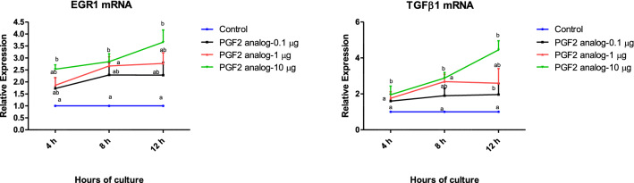
Background: PGF2α is essential for the induction of the corpus luteum regression which in turn reduces progesterone production. Early growth response (EGR) proteins are Cys2-His2-type zinc-finger transcription factor that are strongly linked to cellular proliferation, survival and apoptosis. Rapid elevation of EGR1 was observed after luteolytic dose of PGF2α. EGR1 is involved in the transactivation of many genes, including TGFβ1, which plays an important role during luteal regression.

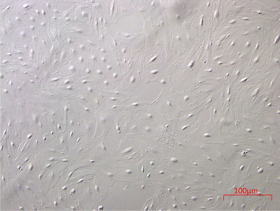
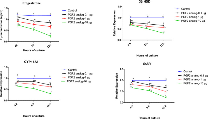
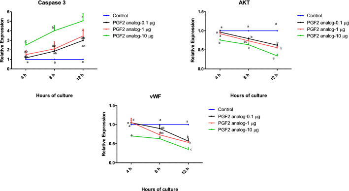
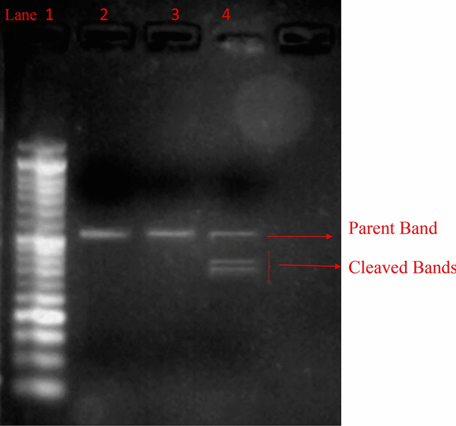
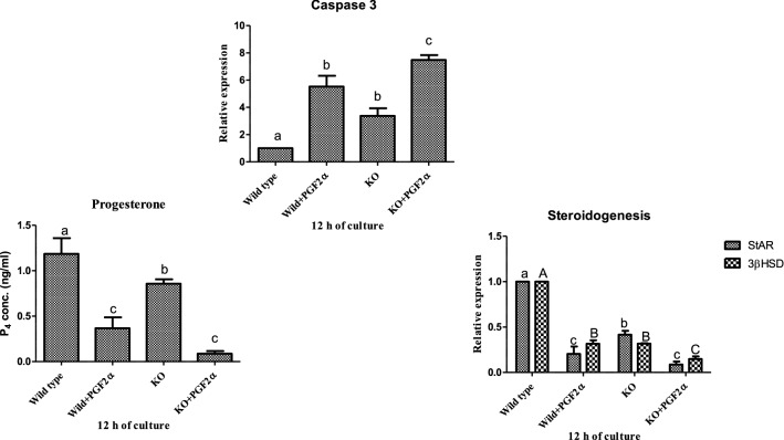
Methods: The current study was conducted in buffalo luteal cells with the aim to better understand the role of EGR1 in transactivation of TGFβ1 during PGF2α induced luteal regression. Luteal cells from mid stage corpus luteum of buffalo were cultured and treated with different doses of PGF2α for different time durations. Relative expression of mRNAs encoding for enzymes within the progesterone biosynthetic pathway (3βHSD, CYP11A1 and StAR); Caspase 3; AKT were analyzed to confirm the occurrence of luteolytic event. To determine if EGR1 is involved in the PGF2α induced luteal regression via induction of TGFβ1 expression, we knocked out the EGR1 gene by using CRISPR/Cas9.

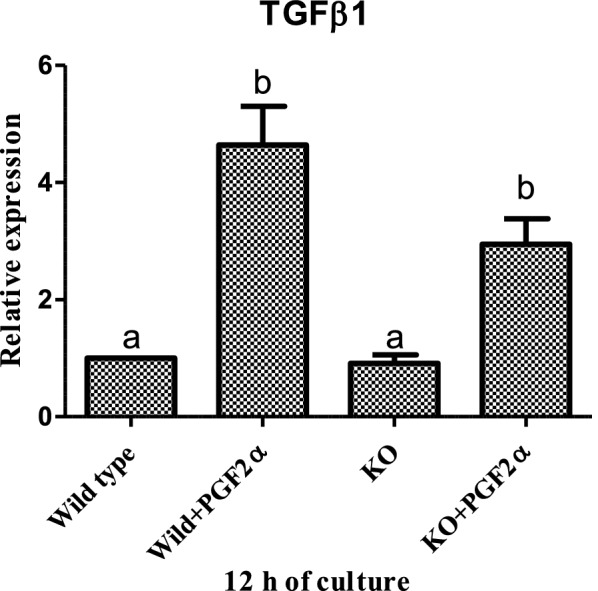
Result: The present experiment determined whether EGR1 protein expression in luteal cells was responsive to PGF2α treatment. Quantification of EGR1 and TGFβ1 mRNA showed significant up regulation in luteal cells of buffalo at 12 h post PGF2α induction. In order to validate the role of PGF2α on stimulating the expression of TGFβ1 by an EGR1 dependent mechanism we knocked out EGR1. The EGR1 ablated luteal cells were stimulated with PGF2α and it was observed that EGR1 KO did not modulate the PGF2α induced expression of TGFβ1. In PGF2α treated EGR1 KO luteal cell, the mRNA expression of Caspase 3 was significantly increased compared to PGF2α treated wild type luteal cells maintained for 12 h. We also studied the influence of EGR1 on steroidogenesis. The EGR1 KO luteal cells with PGF2α treatment showed no substantial difference either in the progesterone concentration or in StAR mRNA expression with PGF2α-treated wild type luteal cells.

Conclusion: These results suggest that EGR1 signaling is not the only factor which plays a role in the regulation of PGF2α induced TGFβ1 signaling for luteolysis.

Keywords: Buffalo; CRISPR/Cas9; Corpus luteum; EGR; Luteolysis.


Conflict of interest statement

The authors declare that they have no competing interests.

Figures

Fig. 1
Fig. 1
Culture of attached luteal cells (×10 magnification)
Fig. 2
Fig. 2
Effect of PGF2α (0.1, 1 and 10 µg/mL) over a period of 4, 8, 12 h (n = 6/condition) on the concentration of progesterone (a) and mRNA expression of 3βHSD (b), CYP11A1 (c) and StAR (d), in in vitro luteal cell culture. 3βHSD 3beta hydroxyl steroid dehydrogenase, CYP11A1 cytochrome P450 side chain cleavage subfamily A1, StAR steroidogenic acute regulatory protein; (p < 0.05), statistically significant
Fig. 3
Fig. 3
Effect of PGF2α (0.1, 1 and 10 µg/mL) over a period of 4, 8, 12 h (n = 6/condition) on mRNA expression of Caspase 3 (a), AKT (b) and vWF (c) in in vitro luteal cell culture. vWF, von Willebrand factor; (p < 0.05), statistically significant
Fig. 4
Fig. 4
Effect of PGF2α (0.1, 1 and 10 µg/mL) over a period of 4, 8, 12 h (n = 6/condition) on mRNA expression of EGR1 (a) and TGFβ1 (b) in in vitro luteal cell culture. EGR1, Early Growth response factor 1; TGF β1 transforming growth factor beta 1; (p < 0.05), statistically significant
Fig. 5
Fig. 5
Gel image of genomic cleavage detection assay of luteal cells transfected with Cas9 and EGR1 SgRNA using Lipofectamine 2000. Lane 1, 50 bp DNA ladder; Lane 2, negative control sample for EGR1 gene; Lane 3 sample without T7/E1 enzyme, Lane 4: sample, showing parent and both the cleaved bands after addition of T7/E1 enzyme
Fig. 6
Fig. 6
Effect of PGF2α (10 µg/mL) on mRNA expression of TGF β1 in MLC and EGR1 KO MLC at 12 h (n = 6/condition). All values are shown as mean ± SEM. Different superscripts denote statistically different values (P < 0.05). EGR 1 KO, Early Growth Response factor 1 Knock out; MLC, Mid stage luteal cells; (p < 0.05), statistically significant
Fig. 7
Fig. 7
Effect of PGF2α (10 µg/mL) on transcriptional abundance of Caspase 3 (a), Progesterone concentration (b) and on transcriptional abundance of StAR, 3βHSD (c) in MLC and EGR1 KO MLC at 12 h (n = 6/condition). All values are shown as mean ± SEM. Different superscripts denote statistically different values (P < 0.05). EGR1 KO Early Growth Response factor 1 Knock out, MLC mid stage luteal cells, StAR steroidogenic acute regulatory protein, 3βHSD 3beta hydroxyl steroid dehydrogenase; (p < 0.05), statistically significant

Similar articles

Cited by

References

    1. Alila HW, Dowd JP, Corradino RA, Harris WV, Hansel W. Control of progesterone production in small and large bovine luteal cells separated by flow cytometry. JRI. 1988;82:645–655. - PubMed
    1. Tsai SJ, Wiltbank MC. Prostaglandin F2α regulates distinct physiological changes in early and mid-cycle bovine corpora lutea. Biol Reprod. 1998;58(2):346–352. doi: 10.1095/biolreprod58.2.346. - DOI - PubMed
    1. Niswender GD, Juengel JL, Silva PJ, Rollyson MK, McIntush EW. Mechanisms controlling the function and life span of the corpus luteum. Physiol Rev. 2000;80(1):1–29. doi: 10.1152/physrev.2000.80.1.1. - DOI - PubMed
    1. Berisha B, Meyer HH, Schams D. Effect of prostaglandin F2 alpha on local luteotropic and angiogenic factors during induced functional luteolysis in the bovine corpus luteum. Biol Reprod. 2010;82(5):940–947. doi: 10.1095/biolreprod.109.076752. - DOI - PubMed
    1. Stocco CO, Lau LF, Gibori G. A calcium/calmodulin-dependent activation of ERK1/2 mediates JunD phosphorylation and induction of nur77 and 20α-hsd genes by prostaglandin F2α in ovarian cells. J Biol Chem. 2002;277(5):3293–3302. doi: 10.1074/jbc.M110936200. - DOI - PubMed

Substances