. 2021 Mar 12;7(11):eabe4983.
doi: 10.1126/sciadv.abe4983. Print 2021 Mar.

Atoh7-independent specification of retinal ganglion cell identity

Affiliations

Atoh7-independent specification of retinal ganglion cell identity

Justin Brodie-Kommit et al. Sci Adv. .

Abstract

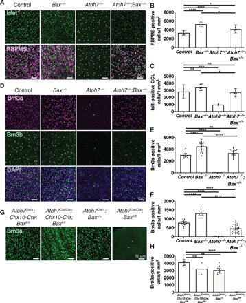
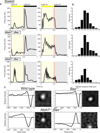
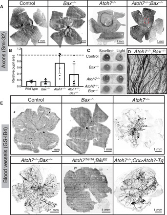
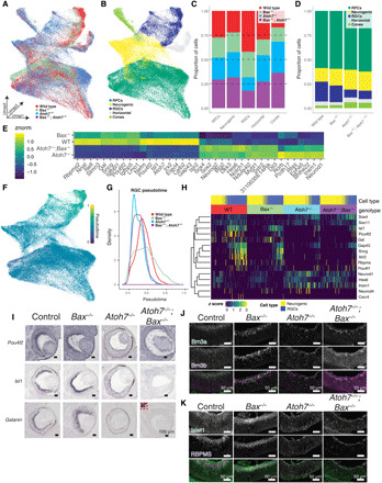
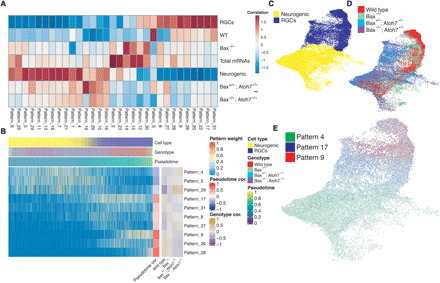
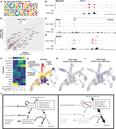
Retinal ganglion cells (RGCs) relay visual information from the eye to the brain. RGCs are the first cell type generated during retinal neurogenesis. Loss of function of the transcription factor Atoh7, expressed in multipotent early neurogenic retinal progenitors leads to a selective and essentially complete loss of RGCs. Therefore, Atoh7 is considered essential for conferring competence on progenitors to generate RGCs. Despite the importance of Atoh7 in RGC specification, we find that inhibiting apoptosis in Atoh7-deficient mice by loss of function of Bax only modestly reduces RGC numbers. Single-cell RNA sequencing of Atoh7;Bax-deficient retinas shows that RGC differentiation is delayed but that the gene expression profile of RGC precursors is grossly normal. Atoh7;Bax-deficient RGCs eventually mature, fire action potentials, and incorporate into retinal circuitry but exhibit severe axonal guidance defects. This study reveals an essential role for Atoh7 in RGC survival and demonstrates Atoh7-dependent and Atoh7-independent mechanisms for RGC specification.


Figures

Fig. 1
Fig. 1. Atoh7-independent development of RGCs.
(A to C) We observed a 25.2 ± 0.9 and 21 ± 3% reduction in RBPMS+ RGC density or Isl1+ GCL cells when comparing Atoh7−/−;Bax−/− to Bax−/− mice. (D to H) Brn3a- and Brn3b-positive RGC density are only moderately reduced when apoptosis is blocked in Atoh7−/−;Bax−/− mice. (G and H) Brn3a-positive RGC numbers are rescued when apoptosis is blocked in all neural RPCs, when Baxlox/lox is crossed to the Chx10-Cre transgene, which is expressed in all RPCs. However, when Bax is specifically removed in Atoh7-Cre knock-in mice, Brn3a RGCs are not rescued. Means ± 95% confidence intervals. Statistical significance tested by one-way analysis of variance (ANOVA) with Tukey’s posttest for multiple comparisons *P < 0.045, **P = 0.0023, ***P < 0.0003, ****P < 0.0001. ns, non-significant.
Fig. 2
Fig. 2. Atoh7 is not required for normal retinal wiring and electrophysiological function.
Cells from Atoh7+/−;Bax−/− and Atoh7−/−;Bax−/− mice responded to light similarly to those from WT. (A) Two different examples, corresponding to two different sets of RGCs per genotype of peristimulus time histogram (PSTH) averaged from 120 repetitions of 1-Hz square-wave flash: WT (top), Atoh7+/−;Bax−/− (middle), and Atoh7−/−;Bax−/− (bottom). (B) Distribution of the spatial receptive field measured using white noise flickering checkerboard: WT (top; cell count = 92), Atoh7+/−;Bax−/− (middle; cell count = 94), and Atoh7−/−;Bax−/− (bottom; cell count = 56). (C) The PSTH of responses to square-wave flash was calculated using 10-ms bins. Mice were assayed at P30. One-way ANOVA, followed by Dunnett’s test, P < 0.05, between Atoh7−/−;Bax−/− and WT.
Fig. 3
Fig. 3. RGC axon guidance and retinal vasculature development require Atoh7-dependent RGCs.
(A and D) Smi-32 labels a subset of RGCs and their axons in an adult WT retina. In Atoh7−/− mice, the Smi-32–positive RGCs have axon guidance deficits. In Atoh7−/−;Bax−/− mice, RGCs have severe axon guidance deficits. Highlighted region (A, Atoh7−/−;Bax−/−) is magnified in (D). (B and C) Using the contralateral PLR as a readout of retina to brain connection allows the appreciation that the severe axon guidance deficits allow for some connection to the brain of the RGCs in the Atoh7−/− or Atoh7−/−;Bax−/− retinas. (E) It has been previously reported that the hyaloid vasculature fails to regress in Atoh7−/− mice, thought to be due to lack of RGCs; however, when the RGC numbers are rescued, in Atoh7−/−;Bax−/−mice, the hyaloid vasculature fails to regress. However, Atoh7 is not necessary for the hyaloid regression and retinal vasculature development, seen using Atoh7tTA/tTA;B&IEE mice, which was previously seen to rescue all of the Atoh7 null phenotypes. When Atoh7 is rescued using the Crx>Atoh7 transgene on the Atoh7 null background, the optic nerve and 12% of RGCs are rescued (22), but the hyaloid vasculature does not regress.
Fig. 4
Fig. 4. Single-cell analysis of E14.5 mutant retinas and gene detection in E14 retinas.
(A and B) Uniform manifold approximation and projection (UMAP) dimension reduction of aggregated E14.5 single-cell dataset colored by (A) genotype and (B) annotated cell type. (C) Proportions of cell types derived from each genotype. (D) Proportions of annotated cell types within each genotype. (E) Heatmap of differentially expressed transcripts across control and Atoh7 knockout (Atoh7−/− or Atoh7−/−;Bax−/−) neurogenic and RGCs. (F) UMAP dimension reduction of cells colored by Scanpy pseudotime values. (G) Density of RGCs along pseudotime by genotype. (H) Heatmap displaying differentially expressed transcripts across the interaction of pseudotime and genotype. Cells are ordered by pseudotime within each genotype. (I) Chromogenic in situ hybridization detecting RNA transcripts of genes from (H). Inset, depicted by red dotted lines, Atoh7−/−;Bax−/− mice in (H) show robust galanin signal in a region outside the retina but minimal signal in the retina (J and K). Immunohistochemistry detecting (J) RGC-specific markers, BRN3A and BRN3B, and (K) pan-RGC markers, ISL1 and RBPMS, in E14 retina from each genotype. Scale bars, 50 μm. RPCs, retinal progenitor cells; RGCs, retinal ganglion cells.
Fig. 5
Fig. 5. scCoGAPS analysis of single-cell dataset and RGC population changes in E12.5 retinas show a developmental delay in Atoh7−/−;Bax−/− mutants.
(A) Heatmap showing the correlation between scCoGAPS pattern and cellular features. (B) Heatmap of pattern weights within individual cells ordered by pseudotime. Pattern correlations with both pseudotime and each genotype are displayed on the right. (C to E) UMAP embedding of single-cell dataset used for scCoGAPS and colored by (C) cell type or (D) genotype. (E) UMAP embedding of dataset and colored by pattern weights of scCoGAPS patterns 4, 17, and 9, displaying progressive pattern usage across RGC development.
Fig. 6
Fig. 6. Cut&Run analysis of Atoh7 genomic binding identifies transcriptional targets of Atoh7.
(A) HOMER motif analysis of most significant de novo (top) enriched motifs within peaks and similarity of enriched motifs to established transcription factor motifs (bottom). (B to D) Atoh7 and IgG Cut&Run sequencing tracks within the (B) Neurod1, (C) Otx2, and (D) Atoh7 genomic loci. Additional E14 ATAC-seq tracks (82) show the alignment of Atoh7 Cut&Run peaks with open chromatin. (E) Average expression of transcripts within the control (WT and Bax−/−) and Atoh7 null (Atoh7−/− or Atoh7−/−;Bax−/−) neurogenic RPCs and RGCs. Transcripts with Atoh7 Cut&Run peaks are indicated in red. Gene names are displayed for transcripts that display both Atoh7 Cut&Run peaks and high residual to the mean. (F) Cellular enrichment of transcript expression within the developmental scRNA-seq dataset (23) of genes with the presence/absence of Atoh7 binding and increased, decreased, or no change in expression within Atoh7 mutant neurogenic RPCs and RGCs compared to control cells. (G to I) UMAP dimension reductions of the developmental scRNA-seq dataset (115) displaying the annotated (G) cell types or (H and I) normalized cellular z scores of (H) Atoh7-bound transcripts with decreased or (I) increased expression in Atoh7 mutants. (J) Model showing the role of Atoh7 in retinal development based on the findings of this study.

Similar articles

Cited by

References

    1. Brown N. L., Patel S., Brzezinski J., Glaser T., Math5 is required for retinal ganglion cell and optic nerve formation. Development 128, 2497–2508 (2001). - PMC - PubMed
    1. Ghiasvand N. M., Rudolph D. D., Mashayekhi M., Brzezinski J. A. IV, Goldman D., Glaser T., Deletion of a remote enhancer near ATOH7 disrupts retinal neurogenesis, causing NCRNA disease. Nat. Neurosci. 14, 578–586 (2011). - PMC - PubMed
    1. Jarman A. P., Grell E. H., Ackerman L., Jan L. Y., Jan Y. N., Atonal is the proneural gene for Drosophila photoreceptors. Nature 369, 398–400 (1994). - PubMed
    1. Khan K., Logan C. V., Kibbin M. M., Sheridan E., Elçioglu N. H., Yenice O., Parry D. A., Fernandez-Fuentes N., Abdelhamed Z. I. A., Al-Maskari A., Poulter J. A., Mohamed M. D., Carr I. M., Morgan J. E., Jafri H., Raashid Y., Taylor G. R., Johnson C. A., Inglehearn C. F., Toomes C., Ali M., Next generation sequencing identifies mutations in Atonal homolog 7 (ATOH7) in families with global eye developmental defects. Hum. Mol. Genet. 21, 776–783 (2012). - PMC - PubMed
    1. Khor C. C., Ramdas W. D., Vithana E. N., Cornes B. K., Sim X., Tay W.-T., Saw S.-M., Zheng Y., Lavanya R., Wu R., Wang J. J., Mitchell P., Uitterlinden A. G., Rivadeneira F., Teo Y.-Y., Chia K.-S., Seielstad M., Hibberd M., Vingerling J. R., Klaver C. C. W., Jansonius N. M., Tai E.-S., Wong T.-Y., van Duijn C. M., Aung T., Genome-wide association studies in Asians confirm the involvement of ATOH7 and TGFBR3, and further identify CARD10 as a novel locus influencing optic disc area. Hum. Mol. Genet. 20, 1864–1872 (2011). - PubMed

Publication types