. 2021 Mar 12;12(3):263.
doi: 10.1038/s41419-021-03527-9.

Evidences for lipid involvement in SARS-CoV-2 cytopathogenesis

Affiliations

Evidences for lipid involvement in SARS-CoV-2 cytopathogenesis

Roberta Nardacci et al. Cell Death Dis. .

Abstract

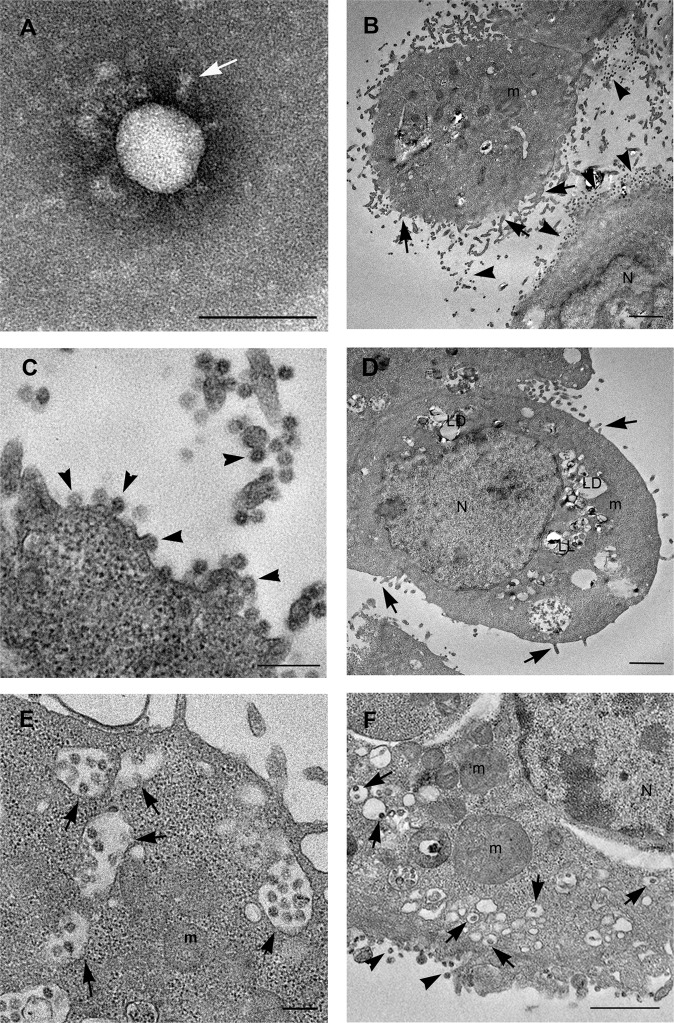
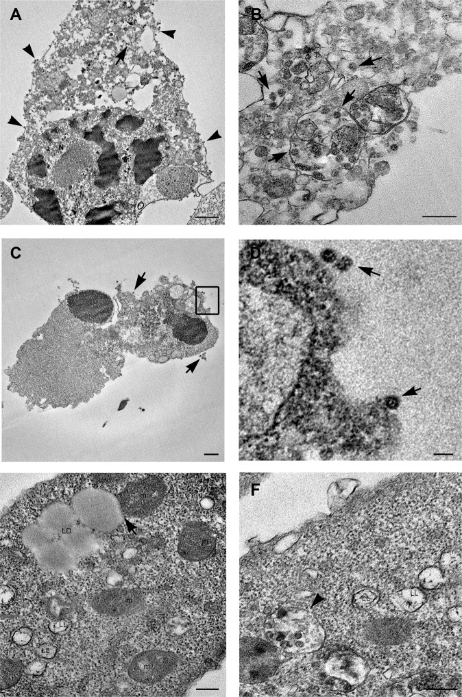
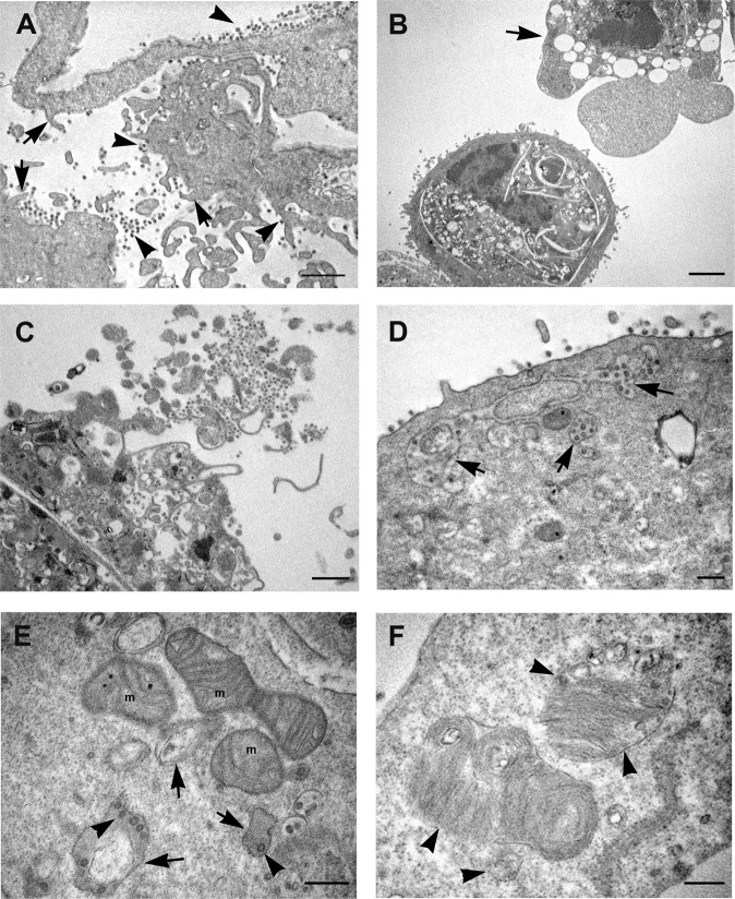
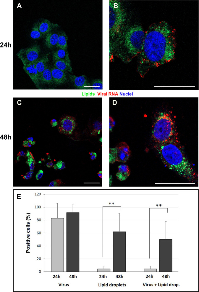
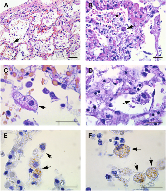
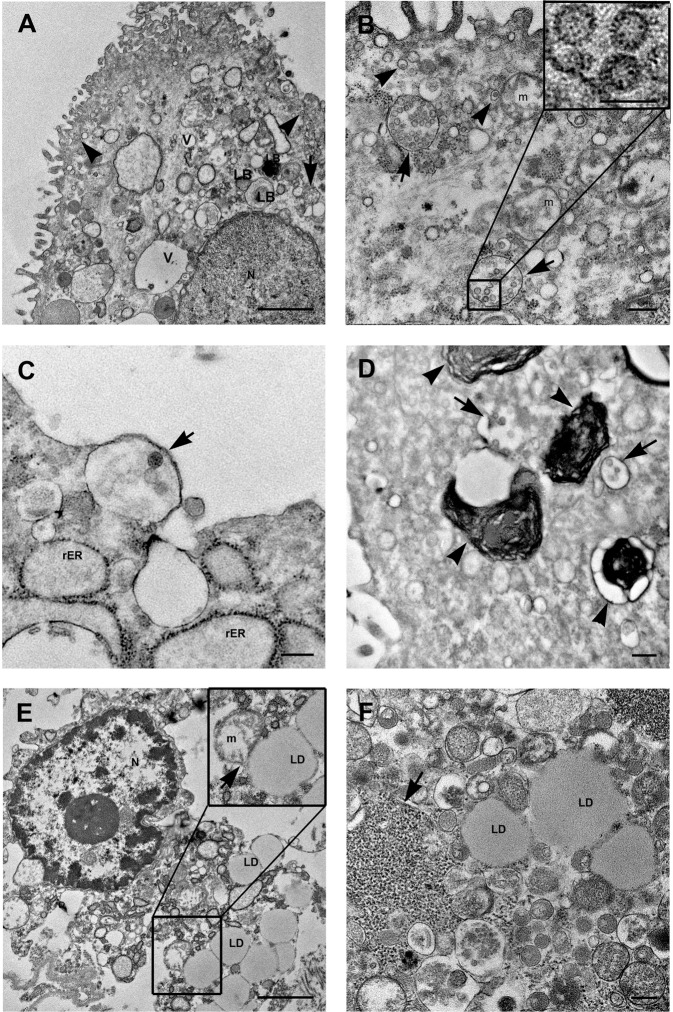
The pathogenesis of SARS-CoV-2 remains to be completely understood, and detailed SARS-CoV-2 cellular cytopathic effects requires definition. We performed a comparative ultrastructural study of SARS-CoV-1 and SARS-CoV-2 infection in Vero E6 cells and in lungs from deceased COVID-19 patients. SARS-CoV-2 induces rapid death associated with profound ultrastructural changes in Vero cells. Type II pneumocytes in lung tissue showed prominent altered features with numerous vacuoles and swollen mitochondria with presence of abundant lipid droplets. The accumulation of lipids was the most striking finding we observed in SARS-CoV-2 infected cells, both in vitro and in the lungs of patients, suggesting that lipids can be involved in SARS-CoV-2 pathogenesis. Considering that in most cases, COVID-19 patients show alteration of blood cholesterol and lipoprotein homeostasis, our findings highlight a peculiar important topic that can suggest new approaches for pharmacological treatment to contrast the pathogenicity of SARS-CoV-2.


Conflict of interest statement

The authors declare no competing interests.

Figures

Fig. 1
Fig. 1. Electron microscopy images of SARS-CoV-2 virus and infected Vero cells.
A Negative staining electron microscopy micrographs of SARS-CoV-2 particle. The virion display a spherical shape, on the surface cone-like shaped spikes are visible (white arrow). BD Infected cells show numerous viruses associated to the plasma membrane (arrowheads) especially found along microvillous projections (arrows). Many lipid droplets (LD) and lipolysosomes (LL) are visible (D). E A great number of vacuoles are present in the cell cytoplasm, many of which contain viral particles (arrows). F Other vesicles, small in size, contain single viral particles, resembled the “spherules” described for other coronaviruses (arrows). Viruses are visible along the plasma membrane (arrowheads). Numerous free ribosomes are diffused in the cell cytosol. N nucleus, m mitochondria, LD lipid droplets, LL lipolysosomes. Scale bars: A = 100 nm; B, D, F = 1 μm; C, E = 200 nm.
Fig. 2
Fig. 2. Electron microscopy features of SARS-CoV-2-infected dying cells.
A, B A dying cell shows an advanced stage of degeneration. The nucleus shows condensed areas and the cytoplasm results empty due to the presence of a high number vacuoles, some of which containing viral particles (arrows). Numerous viral particles are also visible at the cell membrane (arrowheads). C Cell remnants showing viral particles outside of cell (arrows). D Higher magnification of the boxed area visible in C shows viral particles (arrows). E Lipid droplets (LD) are present inside infected cells, some of which are in contact with mitochondria (arrow). Lipolysosomes (LL) with external membrane and whorls are detected. Mitochondria (m) show swollen cristae. F Viral particles in structures resembling lipolysosomes (LL) are visible (arrowhead). N nucleus, m mitochondria, LD lipid droplets, LL lipolysosomes. Scale bars: A, C = 1 um; D = 100 nm; B, E, F = 200 nm.
Fig. 3
Fig. 3. Electron microscopy micrographs of SARS-CoV-1 infected Vero cells.
A Infected cells show a rough aspect, and many microvillous projections are visible all over the cell surface (arrows). Numerous virus particles are visible at the cell membrane or in the extracellular space (arrowheads). B Cytopathic effect of viral infection generates in a number of cells condensation of cytoplasm and apoptotic cell death (arrow). C Cellular lysis with release of cell material with associated virus particles is observed. D Groups of viral particles were found inside vesicles near the plasma membrane of infected cells (arrows). E Morphological modifications of mitochondria (m) are shown, consisting in swelling with a reduction in membrane cristae amount, leading to the formation of vesicles (arrows). Virus particles are visible inside these vesicles (arrowheads). F SARS-CoV-1 infected cells display the presence of multilamellar structure: at the periphery of the structure ribosome-carrying membrane are visible (arrowheads). N nucleus, m mitochondria. Scale bars: AC = 1 μm; DF = 200 nm.
Fig. 4
Fig. 4. Confocal microscopy of SARS-CoV-2-infected Vero cells.
Immunofluorescence dual-labeling detection of viral dsRNA and lipid droplets. A Control cells, display absence of dsRNA signal and diffuse staining with lipophilic dye. B At 24 h post infection some cells showing viral RNA (red) also present lipid droplets (green dots) in the cytoplasm. C, D Numerous lipid droplets are visible in the cell cytoplasms (green dots) of cells 48 h post infection. E The graph shows the percentage of cells displaying viral RNA, lipid droplets staining or both labeling at 24 h and 48 h post infection. Data are mean ± SD from at least three independent experiments, and each experiment included duplicate samples. Statistically significant difference is showed (**p < 0.01). dsRNA (red); lipid droplets (green). Nuclei are stained blue (DAPI). Scale bars: AD = 5 μm.
Fig. 5
Fig. 5. Histopathological changes of lung tissue from COVID-19 patients.
A, B EE staining from lung tissue shows diffuse alveolar damage, with intra-alveolar inflammation, fibrin, and hyaline membranes. Hyperplasia of type II pneumocyte, characterized by amphophilic cytoplasm, large nuclei, and prominent nucleoli is visible (arrows). Necrotic cells are present in the alveolar space (asterisk) (B). C Type II pneumocyte showing signs of degeneration characterized by large nucleus, with fine and uniformly dispersed chromatin and cytoplasm vacuolization (arrow). D Degenerating pneumocytes showing tipical features of pyroptosis (arrow). E, F Immunohistochemistry anti-coronavirus revealed a focal distribution of the positivity (arrows). Scale bars: A = 14 μm; BF = 7 μm.
Fig. 6
Fig. 6. SARS-Cov-2 detecting on lung tissue from COVID-19 patients by transmission electron microscopy.
A, B SARS-CoV-2 particles are detected in type II pneumocytes recognizable by the presence of lamellar bodies, containing surfactant (LB). Viruses are localized in virus containing compartments (arrows). Other vesicles, very small in size, contain single viral particles (arrowheads). Numerous vacuoles (V) are present in the cell cytoplasm. B Mitochondria (m) display swelling with a reduction in membrane cristae amount. Higher magnification of viral particles is visible in the boxed area: black dots are visible inside the viral particles due to cross section through the nucleocapside. C A vesicle containing viral particles (arrow) and enlargement of rough endoplasmic reticulum (rER) are shown. D Lamellar bodies, containing surfactant are visible (arrowheads). Arrows point to virus containing compartments. E, F Abundant lipid droplets (LD) are present inside lung cells. Mitochondria and LDs contacts sites were shown (arrow in insert panel). Free ribosomes are present in the cell cytosol, many of which are compartimentalized (arrow). N nucleus, m mitochondria, rER rough endoplasmic reticulum, LD lipid droplets, LB lamellar bodies, V vacuole. Scale bars: A, E = 1 μm; BD, F = 200 nm; boxed area in B = 100 nm.

Similar articles

Cited by

References

    1. Zhu N, et al. A novel coronavirus from patients with pneumonia in China, 2019. N. Engl. J. Med. 2020;382:727–733. doi: 10.1056/NEJMoa2001017. - DOI - PMC - PubMed
    1. WHO 2020. Coronavirus disease (COVID-19) situation reports. https://www.who.int/emergencies/diseases/novel-coronavirus-2019/situatio... (2020).
    1. Martines RB, et al. Pathology and pathogenesis of SARS-CoV-2 associated with fatal coronavirus disease, United States. Emerg. Infect. Dis. 2020;26:2005–2015. doi: 10.3201/eid2609.202095. - DOI - PMC - PubMed
    1. Shi Y, et al. COVID-19 infection: the perspectives on immune responses. Cell Death Differ. 2020;27:1451–1454. doi: 10.1038/s41418-020-0530-3. - DOI - PMC - PubMed
    1. Celardo I, Pace L, Cifaldi L, Gaudio C, Barnaba V. The immune system view of the coronavirus SARS-CoV-2. Biol. Direct. 2020;15:30. doi: 10.1186/s13062-020-00283-2. - DOI - PMC - PubMed

Publication types

MeSH terms