. 2021 Mar 12;12(1):1654.
doi: 10.1038/s41467-021-21921-x.

ADAR1 RNA editing enzyme regulates R-loop formation and genome stability at telomeres in cancer cells

Affiliations

ADAR1 RNA editing enzyme regulates R-loop formation and genome stability at telomeres in cancer cells

Yusuke Shiromoto et al. Nat Commun. .

Abstract

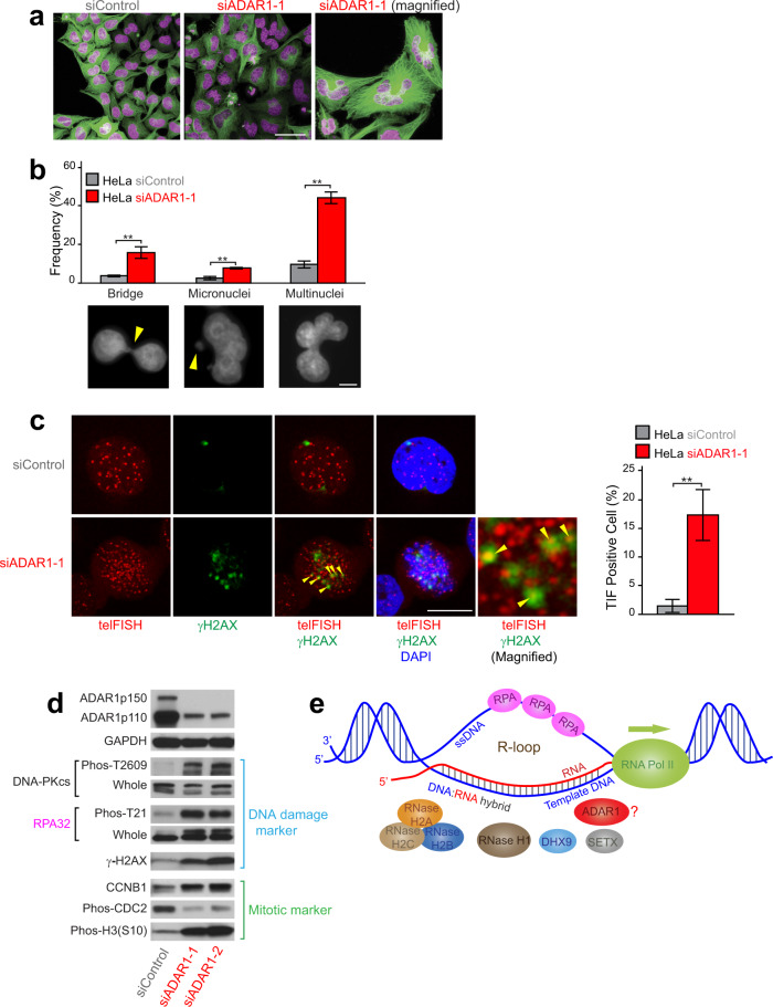
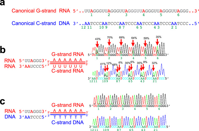
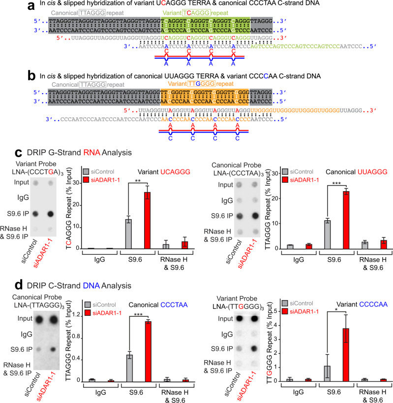
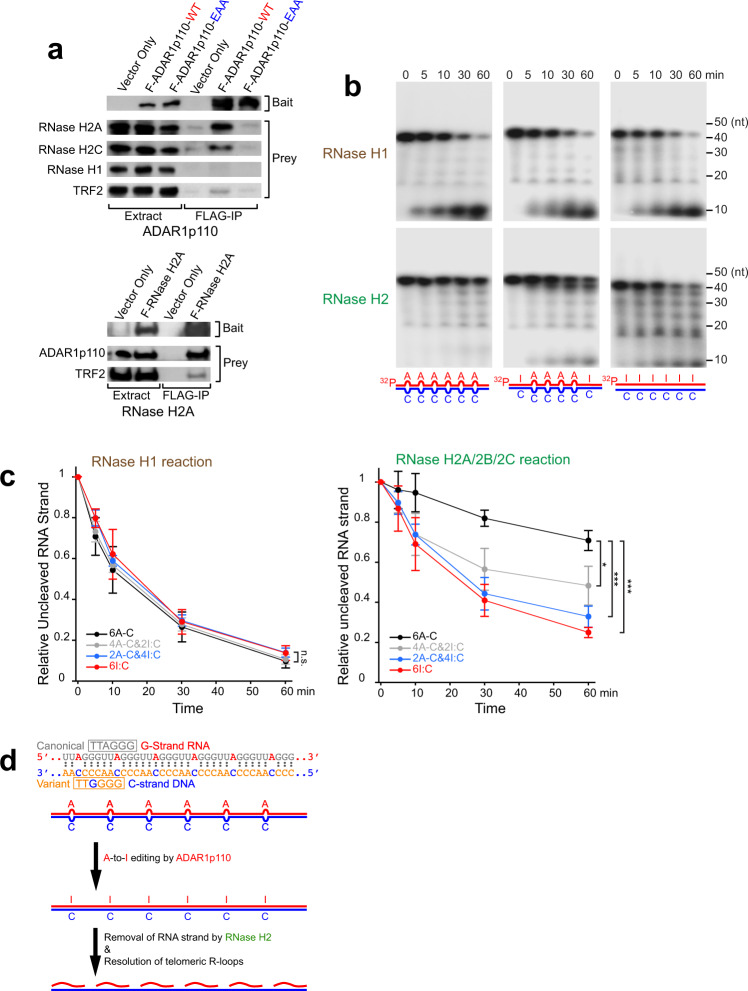
ADAR1 is involved in adenosine-to-inosine RNA editing. The cytoplasmic ADAR1p150 edits 3'UTR double-stranded RNAs and thereby suppresses induction of interferons. Loss of this ADAR1p150 function underlies the embryonic lethality of Adar1 null mice, pathogenesis of the severe autoimmune disease Aicardi-Goutières syndrome, and the resistance developed in cancers to immune checkpoint blockade. In contrast, the biological functions of the nuclear-localized ADAR1p110 remain largely unknown. Here, we report that ADAR1p110 regulates R-loop formation and genome stability at telomeres in cancer cells carrying non-canonical variants of telomeric repeats. ADAR1p110 edits the A-C mismatches within RNA:DNA hybrids formed between canonical and non-canonical variant repeats. Editing of A-C mismatches to I:C matched pairs facilitates resolution of telomeric R-loops by RNase H2. This ADAR1p110-dependent control of telomeric R-loops is required for continued proliferation of telomerase-reactivated cancer cells, revealing the pro-oncogenic nature of ADAR1p110 and identifying ADAR1 as a promising therapeutic target of telomerase positive cancers.


Conflict of interest statement

The authors declare no competing interests.

Figures

Fig. 1
Fig. 1. ADAR1 depletion resulted in abnormalities of the nucleus and upregulation of DNA damage pathway and cell cycle marker genes.
a HeLa cells were first transfected with siControl or siADAR1 (siADAR1-1) for 72 h and then treated with CellLight Tubulin-GFP. Nuclei were visualized by staining of DNA with SiR-DNA reagent. Representative images taken from real-time videos (Supplementary Movies S1 and 2) are presented. Scale bar, 50 μm. b The frequency of abnormalities of the nucleus (nucleoplasmic bridge, micronuclei, and multinuclei) was estimated by examining at least 200 individual HeLa cells treated with siControl or siADAR1-1 RNAs. Values are mean ± standard error (n = 3, biologically independent samples) with significant differences by two-tailed Student’s t test indicated, **P < 0.01. Scale bar, 5 μm. c Telomere DNA damages in ADAR1-depleted cells. Telomere FISH and immunostaining for γH2AX revealed significantly increased telomere dysfunction-induced foci (TIF, indicated by yellow arrowheads), suggesting the causative relevance of the telomeric repeat DNA damage to chromosome abnormality detected in ADAR1-depleted HeLa cells. At least 200 individual HeLa cells treated with siControl or siADAR1-1 RNAs were examined. HeLa cells with one or more TIFs were counted as TIF-positive cells. Values are mean ± SD (n = 3, biologically independent samples) with significant differences by two-tailed Student’s t test indicated, **P < 0.01. Scale bar, 10 μm. b, c All individual experimental data values and exact P values are presented in Source Data file. d Western blotting analysis was done using total cell extracts from HeLa cells treated with siControl or two separate siADAR1 (siADAR1-1 and -2) RNAs for 72 h. Protein molecular weight markers are presented in Source Data file. e The R-loop structure consisting of an RNA:DNA hybrid formed between the RNA strand newly transcribed by RNA polymerase II and the template DNA strand with the single-stranded antisense DNA bound with ssDNA-binding protein RPA as well as major regulators are schematically shown.
Fig. 2
Fig. 2. A-to-I editing activity of ADAR1p110, not ADAR1p150 or ADAR2, is required for suppression of R-loops.
ae Dot blot analysis for RNA:DNA hybrids was conducted using control oligos (a) or genomic DNA (b–e). a The S9.6 antibody recognized specifically RNA:DNA but not DNA:DNA or RNA:RNA oligo duplex controls. b, c Increased RNA:DNA hybrids were detected only in ADAR1-depleted but not in ADAR2-depleted HeLa cells. b The S9.6 antibody signals were abolished by E. coli-RNase H treatment, confirming specific detection of RNA:DNA hybrids. d Comparison of RNA:DNA hybrid levels between depletion of ADAR1 versus depletion of known R-loop regulators. e Increased RNA:DNA hybrid formation resulting from depletion of endogenous ADAR1 was rescued by infection of ADAR1p110-WT (wild type) but not by infection of ADAR1p110-E912A deamination defective mutant or ADAR1p150-WT. ce Data are mean ± SD (n = 3, biological replicates); significant differences were identified by two-tailed Student’s t tests: *P < 0.05, **P < 0.01, ***P < 0.001, n.s., not significant. All individual experimental data values and exact P values are presented in Source Data file.
Fig. 3
Fig. 3. Accumulation of R-loops at telomeres in ADAR1-depleted cells.
a ADAR1 depletion had no effects on already known sites prone to the formation of R-loops. Six sites were examined by qPCR analysis of DRIP products. PCR primers used are listed in Supplementary Data 1. Data are mean ± SD (n = 3, biological replicates); significant differences were identified by two-tailed Student’s t tests: n.s., not significant. b DRIP products were subjected to genomic DNA dot blot hybridization analysis with a probe containing the G-rich-telomere canonical repeat (TTAGGG), or a probe for α-satellite repeat, Alu, or LINE1 consensus sequence (Supplementary Data 1). ADAR1 depletion resulted in increased the formation of RNA:DNA hybrids specifically at telomeric repeats, which was abolished by E. coli-RNase H treatment prior to DRIP. c Significance of the increased R-loop formation at telomeric repeats was confirmed by conducting three independent dot blot hybridization analyses of DRIP products. Data are mean ± SD (n = 3, biological replicates); significant differences were identified by two-tailed Student’s t tests: **P < 0.01, n.s., not significant. a, c All individual experimental data values and exact P values are presented in Source Data file.
Fig. 4
Fig. 4. ADAR1p110 cannot edit completely matched RNA:DNA hybrids carrying telomeric repeat sequences.
a Canonical telomeric repeat sequences of G-strand RNA (red) and C-strand DNA (blue) are shown. Six adenosines of the G-strand RNA and twelve adenosines of the C-strand DNA are indicated by numbers 1–6 and 1–12, respectively. b In vitro editing assay for completely matched telomeric repeat dsRNA was conducted using ADAR1p110-WT recombinant protein. c In vitro editing assay for completely matched telomeric repeat RNA:DNA hybrids using ADAR1p110-WT recombinant protein. No significant levels of editing for matched RNA:DNA hybrids were detected. b, c PCR products (RT-PCR-amplified RNA strands and PCR-amplified DNA strands) were subjected to Sanger sequencing. The editing frequency was estimated as the % ratio of the guanosine (black) peak over the sum of guanosine and adenosine (green) peaks of the sequencing chromatograms. Editing frequency estimated for three independent experiments (n = 3, technical replicates) is presented in Supplemental information (Supplementary Data 2 and Source Data file).
Fig. 5
Fig. 5. Detection of telomeric variant repeats in ALT-positive and non-ALT cancer cell lines.
a Dot blot hybridization analysis for genomic DNA samples was conducted using three separate telomeric repeat probes capable of distinguishing a single-nucleotide mismatch (Supplementary Fig. 4a, b). In addition to canonical TTAGGG signals, varying amounts of TCAGGG and TTGGGG variant repeat signals were detected in both ALT and non-ALT cancer cell lines, but not in primary human fibroblast cells. b Quantitation of canonical and variant telomeric repeats was done by comparing dot blot signals of genomic DNA and canonical and variant repeat-specific control oligos. Three independent dot blot hybridization analyses were performed. Data are mean ± SD (n = 3, biological replicates). All individual experimental data values are presented in Source Data file.
Fig. 6
Fig. 6. Increased telomeric RNA:DNA hybrids containing variant repeats in ADAR1-depleted cells.
Formation of telomeric repeat RNA:DNA hybrids containing A–C mismatches by in cis slipped hybridization (a, b). a TERRA RNAs transcribed from the region containing four TCAGGG (green) variant repeats surrounded by TTAGGG (gray) canonical repeats form an RNA:DNA hybrid containing four C–A mismatches by in cis slipped hybridization to the C-strand DNA containing canonical TTAGGG (CCCTAA) repeats. b TERRA RNAs transcribed from the region containing four TTGGGG (orange) variant repeats surrounded by TTAGGG (gray) canonical repeats form an RNA:DNA hybrid containing four A–C mismatches by in cis slipped hybridization to the C-strand DNA containing TTGGGG (CCCCAA) variant repeats. c, d Detection of increased RNA:DNA hybrids containing TCAGGG and TTGGGG variant repeats in ADAR1-depleted HeLa cells. c DRIP products were examined for G-strand RNAs of UCAGGG variant and UUAGGG canonical repeats by dot blot analysis using high-affinity LNA-oligonucleotide probes capable of distinguishing a single-nucleotide mismatch (Supplementary Fig. 4c). d Similarly, DRIP products were examined for C-strand DNAs of TTAGGG (CCCTAA) canonical and TTGGGG (CCCCAA) variant repeats using LNA-oligonucleotide probes capable of distinguishing a single-nucleotide mismatch (Supplementary Fig. 4d). c, d Dot blot signals were abolished by E. coli-RNase H treatment prior to DRIP. The significance of the increase in RNA:DNA hybrids containing telomeric canonical and variant repeats (RNA and DNA strands) was confirmed by conducting three independent dot blot hybridization analysis of DRIP products. Data are mean ± SD (n = 3, biological replicates); significant differences were identified by two-tailed Student’s t tests: *P < 0.05, **P < 0.01, ***P < 0.001. All individual experimental data values and exact P values are presented in Source Data file.
Fig. 7
Fig. 7. ADAR1p110 edits both RNA and DNA strands of telomeric repeat RNA:DNA hybrids containing A–C mismatches.
ac In vitro editing assay for telomeric repeat dsRNA (a) and RNA:DNA hybrids (b, c) containing A–C or C–A mismatches was conducted using ADAR1p110-WT recombinant protein. PCR products (RT-PCR-amplified RNA strands and PCR-amplified DNA strands) were subjected to Sanger sequencing. Editing frequency estimated for three independent experiments (n = 3, technical replicates) is presented in Supplemental information (Supplementary Data 2 and Source Data file).
Fig. 8
Fig. 8. Editing of A–C mismatches in telomeric repeat RNA:DNA hybrids facilitates RNase H2 cleavage of RNA strands.
a F-ADAR1p110-WT recombinant protein was copurified with endogenous RNase H2 subunits H2A and H2C, but not with RNase H1. No RNase H2 association detected with a dsRNA-binding defective mutant F-ADAR1p110-EAA (upper panels). Similarly, FLAG-RNase H2A pulled down endogenous ADAR1p110 (lower panels). Note that both FLAG-AFAR1p110 and FLAG-RNase H2A pulled down endogenous TRF2, a telomere-binding protein and a member of the Shelterin complex, supporting our hypothesis that ADAR1 and RNase H2 collaborate together to resolve telomeric R-loops. Protein molecular weight markers are presented in Source Data file. b In vitro assay for digestion of 5′-32P-labeled RNA strands of telomeric repeat RNA:DNA hybrids by RNase H1 or RNase H2A/2B/2C complexes. RNase H2 could not degrade the RNA strands of telomeric repeat RNA:DNA hybrids containing six A–C mismatches, but began digesting RNA strands of RNA:DNA hybrids containing four A–C mismatches. Replacement of all A–C mismatches with I:C-matched base pairs resulted in efficient digestion of RNA strands by RNase H2. RNase H1 degraded RNA strands regardless of the number of A–C mismatches. c Time course of digestion of RNA strands of telomeric repeat RNA:DNA hybrids with varying numbers of A–C mismatches by RNase H1 or RNase H2. Data are mean ± SD (n = 4, technical replicates); significant differences were identified by two-tailed Student’s t tests: *P < 0.05, ***P < 0.001, n.s., not significant. All individual experimental data values and exact P values are presented in Supplementary Data 3. d A-to-I editing of A–C mismatches to I:C base pairs in RNA:DNA hybrids facilitates digestion of RNA strands by RNase H2.
Fig. 9
Fig. 9. ADAR1 regulates accumulation of telomeric R-loops only in non-ALT cells.
a Detection of increased RNA:DNA hybrids only in non-ALT cells. Genomic DNA samples (0.25 µg) collected from various cells treated with siControl or siADAR1-1 RNAs for 72 h were examined by dot blot assay using the S9.6 antibody. Data are mean ± SD (n = 3, biological replicates); significant differences were identified by two-tailed Student’s t tests: *P < 0.05, **P < 0.01, ***P < 0.001, n.s., not significant. b DRIP products from select cell lines were further subjected to dot blot analysis for the C-strand telomeric repeat DNA using the canonical telomeric repeat (TTAGGG) specific probe (Supplementary Fig. 4a). Telomeric repeat RNA:DNA hybrids were detected only in HEK293T and HCT116 non-ALT cancer cell lines. Data are mean ± SD (n = 3, biological replicates); significant differences were identified by two-tailed Student’s t tests: *P < 0.05, **P < 0.01, n.s., not significant. a, b All individual experimental data values and exact P values are presented in Source Data file.
Fig. 10
Fig. 10. M-phase-specific interaction of ADAR1p110 and RNase H2 in non-ALT cancer cells.
a Elevated ADAR1 and RNase H2A expression levels detected in non-ALT cells. Western blotting analysis was performed using total cell extract proteins and specific antibodies (Supplementary Data 4). b Interaction between ADAR1p110 and RNase H2 detected only in non-ALT cancer cells. FLAG-ADAR1p110-WT recombinant protein was copurified with endogenous RNase H2 subunit H2A only from non-ALT cell lines. c Elevated RNase H2A expression levels and also increased interaction of ADAR1p110 with RNase H2A detected specifically at M phase. FLAG-ADAR1p110-WT pull-down experiments were conducted using HEK293T cells synchronized at M phase using the thymidine-nocodazole double block system. ac Protein molecular weight markers are presented in Source Data file.

References

    1. Eisenberg E, Levanon EY. A-to-I RNA editing - immune protector and transcriptome diversifier. Nat. Rev. 2018;19:473–490. doi: 10.1038/s41576-018-0006-1. - DOI - PubMed
    1. Nishikura K. A-to-I editing of coding and non-coding RNAs by ADARs. Nat. Rev. Mol. Cell Biol. 2016;17:83–96. doi: 10.1038/nrm.2015.4. - DOI - PMC - PubMed
    1. Walkley CR, Li JB. Rewriting the transcriptome: adenosine-to-inosine RNA editing by ADARs. Genome Biol. 2017;18:205. doi: 10.1186/s13059-017-1347-3. - DOI - PMC - PubMed
    1. Mannion N, Arieti F, Gallo A, Keegan LP, O’Connell MA. New insights into the biological role of mammalian ADARs; the RNA editing proteins. Biomolecules. 2015;5:2338–2362. doi: 10.3390/biom5042338. - DOI - PMC - PubMed
    1. Samuel CE. Adenosine deaminase acting on RNA (ADAR1), a suppressor of double-stranded RNA-triggered innate immune responses. J. Biol. Chem. 2019;294:1710–1720. doi: 10.1074/jbc.TM118.004166. - DOI - PMC - PubMed

Publication types