. 2022 Aug;42(6):1897-1908.
doi: 10.1007/s10571-021-01071-w. Epub 2021 Mar 13.

Pericytes Regulate Cerebral Perfusion through VEGFR1 in Ischemic Stroke

Affiliations

Pericytes Regulate Cerebral Perfusion through VEGFR1 in Ischemic Stroke

Chang-Xiong Gong et al. Cell Mol Neurobiol. 2022 Aug.

Abstract

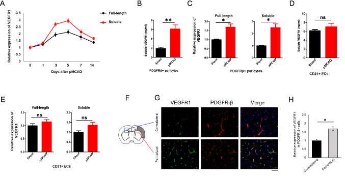
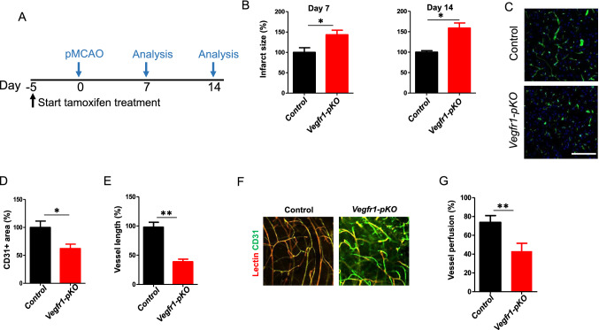
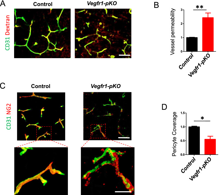
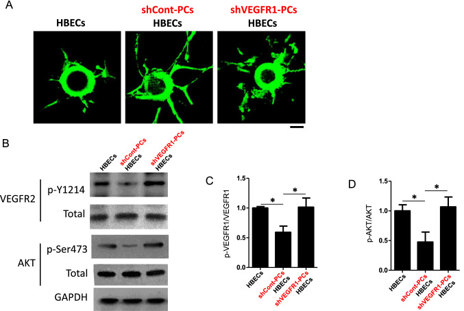
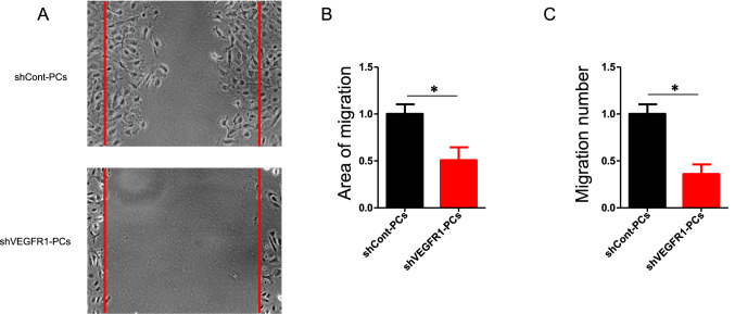
Neurons in the penumbra (the area surrounding ischemic tissue that consists of still viable tissue but with reduced blood flow and oxygen transport) may be rescued following stroke if adequate perfusion is restored in time. It has been speculated that post-stroke angiogenesis in the penumbra can reduce damage caused by ischemia. However, the mechanism for neovasculature formation in the brain remains unclear and vascular-targeted therapies for brain ischemia remain suboptimal. Here, we show that VEGFR1 was highly upregulated in pericytes after stroke. Knockdown of VEGFR1 in pericytes led to increased infarct area and compromised post-ischemia vessel formation. Furthermore, in vitro studies confirmed a critical role for pericyte-derived VEGFR1 in both endothelial tube formation and pericyte migration. Interestingly, our results show that pericyte-derived VEGFR1 has opposite effects on Akt activity in endothelial cells and pericytes. Collectively, these results indicate that pericyte-specific expression of VEGFR1 modulates ischemia-induced vessel formation and vascular integrity in the brain.

Keywords: Cerebrovascular; Ischemia; Pericyte; Stroke; VEGFR1.


Conflict of interest statement

The authors have no conflict of interest to declare.

Figures

Fig. 1
Fig. 1
Expression of VEGFR1 in pericyte is increased after pMCAO. a qPCR analysis of full-length and soluble VEGFR1 in ischemic tissue at indicated days after pMCAO (n = 5 mice). Error bars: s.e.m. b Soluble VEGFR1 in PDGFRβ+cell culture supernatants was determined by ELISA (n = 5 mice). Error bars: s.e.m. ** P < 0.01. c qPCR analysis of full-length and soluble VEGFR1 in PDGFRβ+pericytes sorted from pMCAO or sham group at day 7 (n = 5 mice). Error bars: s.e.m. * P < 0.05. d Soluble VEGFR1 in CD31+ cell culture supernatants was determined by ELISA (n = 5 mice). Error bars: s.e.m. ns = not significant. e qPCR analysis of full-length and soluble VEGFR1 in CD31 + ECs sorted from pMCAO or sham group at day 7 (n = 5 mice). Error bars: s.e.m. ns = not significant. f Schematic of the pMCAO model. The shaded area indicates the infarct area. The framed areas indicate the peri-infarct zones in contralateral (blue) and ischemic (red) hemisphere. g VEGFR1 staining of contralateral and peri-infarct area from pMCAO brains at day 7. Scale bar, 50 μm. h Quantitation of VEGFR1 expression in the contralateral and peri-infarct area. Error bars, s.e.m. *P < 0.05
Fig. 2
Fig. 2
Reduced post-ischemia neovascularization in Vegfr1-pKO mice. a Experimental scheme of tamoxifen administration for the generation of Vegfr1-pKO mice. b the infarct area in Vegfr1-pKO mice relative to control at 7 days and 14 days after pMCAO. (n = 5 mice). Error bars: s.e.m. *P < 0.05. c CD31 staining of peri-infarct area at Day 7 post ischemia induction by pMCAO. Scale bar, 50 µm. d Quantification of CD31 + area at Day 7 presented as relative values in comparison to control mice. *p < 0.05, n = 5. e Vessel length. Error bars: s.e.m. **p < 0.01, n = 5. f Lectin perfusion (red) into peri-infarct vessels (green) was determined. Yellow color (green/red double-staining) indicates blood perfused vessels. Scale bar, 50 µm. g Quantification of vessel perfusion % = lectin + CD31 + area/total CD31 + area × 100, **p < 0.01, n = 5
Fig. 3
Fig. 3
Impaired vascular integrity and pericyte coverage in pericyte-specific VEGFR1 deficiency. a Dextran (red) was injected to mice before euthanization 7 days post pMCAO. Brain tissue from control and Vegfr1-pKO mice was stained for CD31 (green) to label the blood vessels. Scale bar, 50 μm. b Quantification of the ratio of dextran outside vs inside of blood vessel. Error bars: s.e.m. **p < 0.01, n = 5. c Representative confocal microscopy three dimensional reconstruction images of brain sections showing NG2-positive pericyte coverage (red) and CD31-positive endothelial cells (green) in the peri-infarct regions of control and Vegfr1-pKO mice. Higher magnification showed disrupted pericytes-endothelial cells association in Vegfr1-pKO mice. scale bars, 50 μm; inset, 20 μm. d Quantification of pericyte coverage of blood vessels. n = 4 per group
Fig. 4
Fig. 4
VEGFR1 deletion exert differential effects on VAkt activation in ECs and pericytes. PDGFβ + cells and CD31 + cells were sorted from control and Vegfr1-pKO mice at 7 days after pMCAO, respectively. a–e Analyses of VEGFR2 (Y-1214) and Akt (Ser473) phosphorylation and VEGFR1 (full-length, soluble) expression in ECs (n = 5 mice). Error bars: s.e.m. * P < 0.05, **p < 0.01, ns = not significant. f–j Analyses of VEGFR2 (Y-1214) and Akt (Ser473) phosphorylation and VEGFR1 (full-length, soluble) expression in pericytes (n = 5 mice). Error bars: s.e.m. *P < 0.05, **p < 0.01, ns = not significant
Fig. 5
Fig. 5
Pericytes regulate brain endothelial tube formation through paracrine VEGFR1 pathway. a Endothelial sprouts extending from HBECs-coated beads in 3D fibrin gel culture were imaged at day 5. Scale bar, 50 µm. b–d Western blot analysis of VEGFR2 (Y-1214) and Akt (Ser473) phosphorylation in HBECs that cultured in indicated conditions. HBECs were harvested and sorted after 5 days culturing in the gel. n = 3. Error bars: s.e.m. *P < 0.05
Fig. 6
Fig. 6
VEGFR1 is required for pericyte migration. a shCont-PCs and shVEGFR1-PCs were cultured on fibronectin until reaching confluence. A uniform linear scrape wound was made across the cell layer. Three independent experiments were performed. b Area of migration analyzed 24 h after making scratches (n = 5). Error bars: s.e.m. *P < 0.05. c shCont-PCs or shVEGFR1-PCs were seeded in the upper chambers of transwell inserts, parental HBECs were seeded in the lower chambers. After 4 h, cells on the lower side of the filter were stained with 0.5% crystal violet and scored in five independent fields (n = 3). Error bars: s.e.m. *P < 0.05
Fig. 7
Fig. 7
Membrane-localized VEGFR1 regulates Akt signaling in pericytes. a Immunocytochemical analysis of VEGFR1 localization identified prominent nuclear VEGFR1 in cultured pericytes upon PlGF stimulation. b–c Western blot analysis of Akt (Ser473) phosphorylation in shCont-PCs and shVEGFR1-PCs. n = 3. Error bars: s.e.m. **p < 0.01

References

    1. Al Ahmad A, Taboada CB, Gassmann M, Ogunshola OO (2011) Astrocytes and pericytes differentially modulate blood-brain barrier characteristics during development and hypoxic insult. J Cereb Blood Flow Metab 31(2):693–705 - PMC - PubMed
    1. Arenillas JF, Sobrino T, Castillo J, Davalos A (2007) The role of angiogenesis in damage and recovery from ischemic stroke. Curr Treat Options Cardiovasc Med 9(3):205–212 - PubMed
    1. Blanco R, Gerhardt H (2013) VEGF and Notch in tip and stalk cell selection. Cold Spring Harb Perspect Med 3(1):a006569 - PMC - PubMed
    1. Boucher JM, Clark RP, Chong DC, Citrin KM, Wylie LA, Bautch VL (2017) Dynamic alterations in decoy VEGF receptor-1 stability regulate angiogenesis. Nat Commun 8:15699 - PMC - PubMed
    1. Carmeliet P, Jain RK (2011) Molecular mechanisms and clinical applications of angiogenesis. Nature 473(7347):298–307 - PMC - PubMed

LinkOut - more resources