. 2021 Aug;231(4):1478-1495.
doi: 10.1111/nph.17338. Epub 2021 Jun 10.

Functional understanding of secondary cell wall cellulose synthases in Populus trichocarpa via the Cas9/gRNA-induced gene knockouts

Affiliations

Functional understanding of secondary cell wall cellulose synthases in Populus trichocarpa via the Cas9/gRNA-induced gene knockouts

Wenjing Xu et al. New Phytol. 2021 Aug.

Abstract

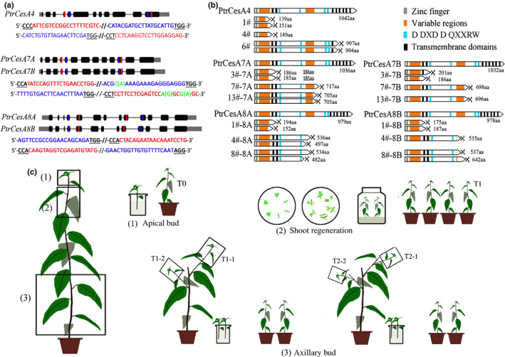
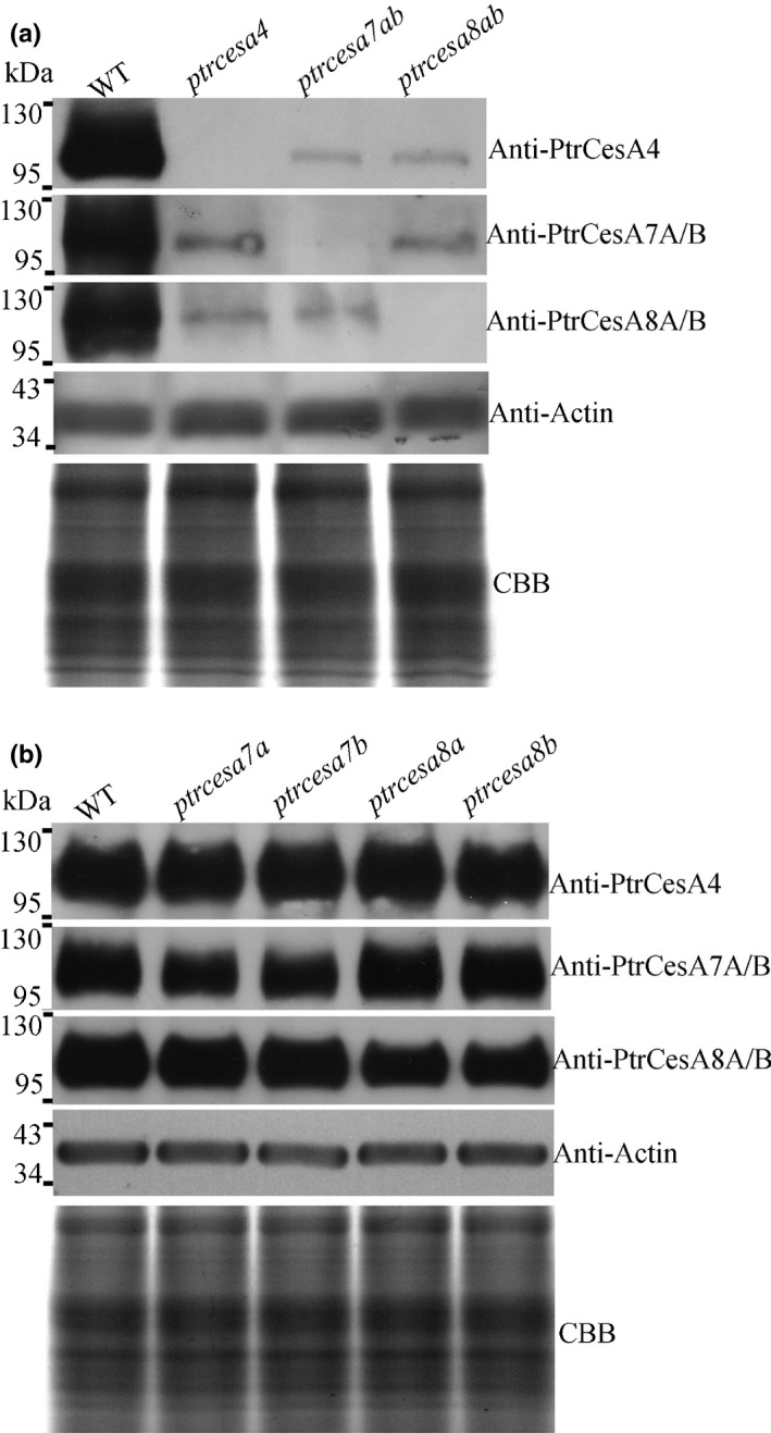
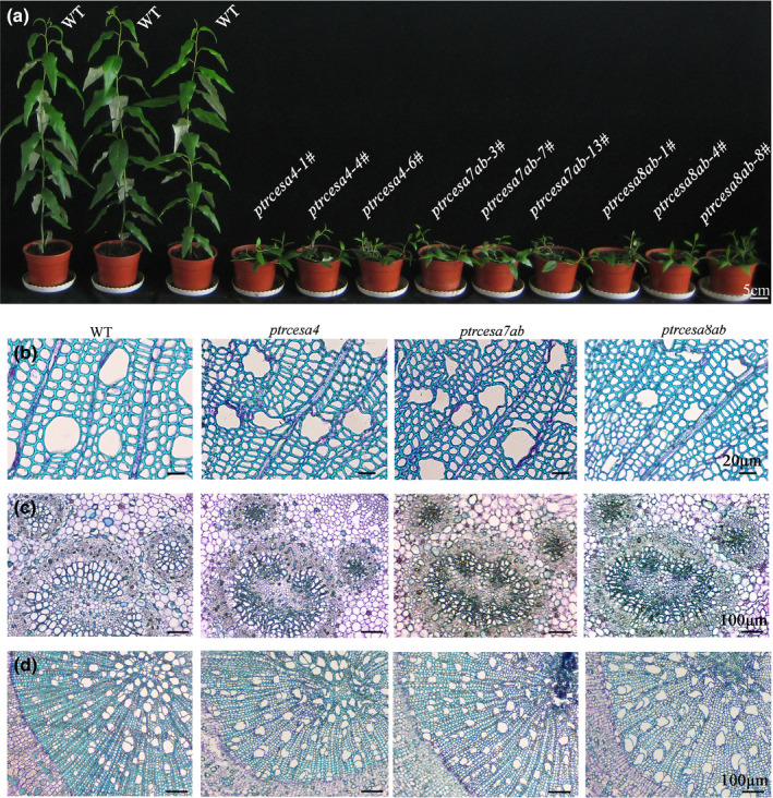
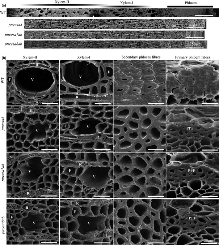
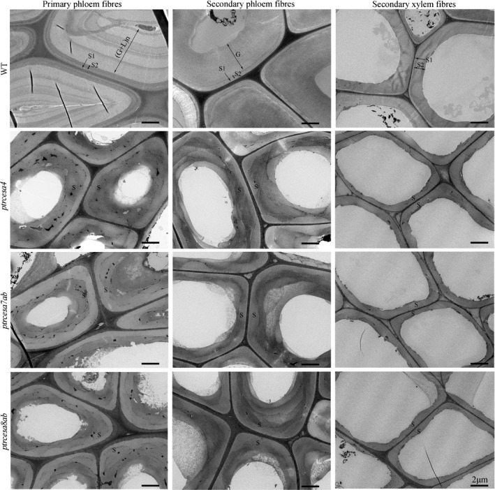
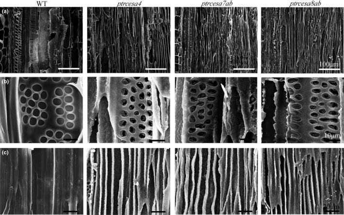
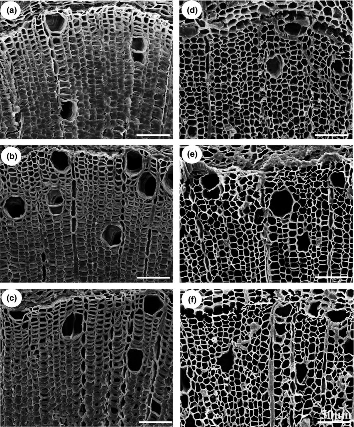
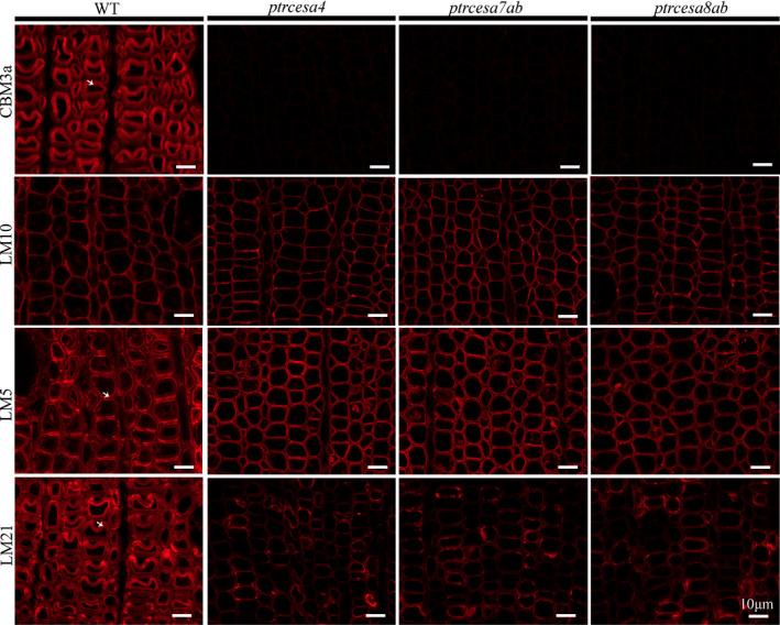
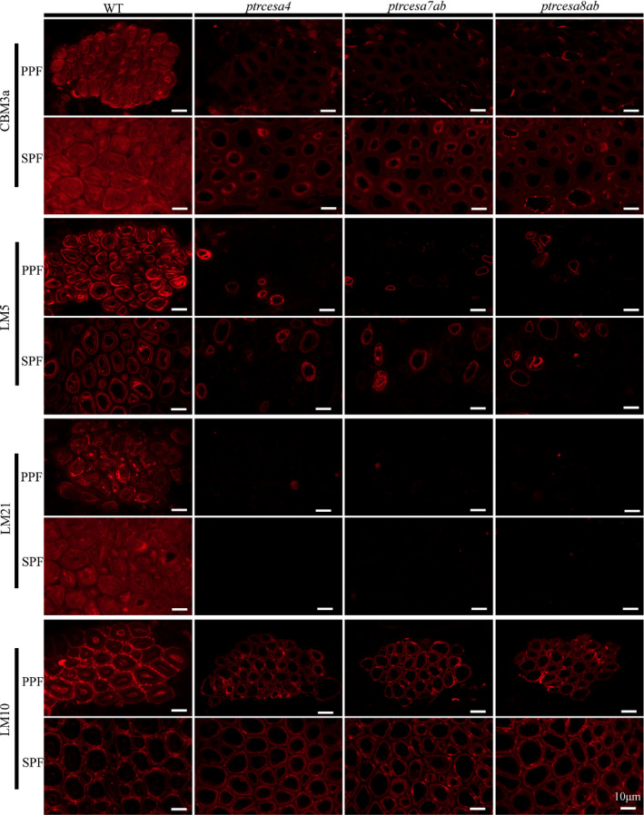
Plant cellulose is synthesized by a large plasma membrane-localized cellulose synthase (CesA) complex. However, an overall functional determination of secondary cell wall (SCW) CesAs is still lacking in trees, especially one based on gene knockouts. Here, the Cas9/gRNA-induced knockouts of PtrCesA4, 7A, 7B, 8A and 8B genes were produced in Populus trichocarpa. Based on anatomical, immunohistochemical and wood composition evidence, we gained a comprehensive understanding of five SCW PtrCesAs at the genetic level. Complete loss of PtrCesA4, 7A/B or 8A/B led to similar morphological abnormalities, indicating similar and nonredundant genetic functions. The absence of the gelatinous (G) layer, one-layer-walled fibres and a 90% decrease in cellulose in these mutant woods revealed that the three classes of SCW PtrCesAs are essential for multilayered SCW structure and wood G-fibre. In addition, the mutant primary and secondary phloem fibres lost the n(G + L)- and G-layers and retained the thicker S-layers (L, lignified; S, secondary). Together with polysaccharide immunolocalization data, these findings suggest differences in the role of SCW PtrCesAs-synthesized cellulose in wood and phloem fibre wall structures. Overall, this functional understanding of the SCW PtrCesAs provides further insights into the impact of lacking cellulose biosynthesis on growth, SCW, wood G-fibre and phloem fibre wall structures in the tree.

Keywords: Cas9; Populus trichocarpa; cellulose synthase (CesA); gRNA; gelatinous layer (G-layer); gene knockout; phloem fibre; secondary cell wall (SCW); tension wood.


Figures

Fig. 1
Fig. 1
Cas9/gRNA‐induced mutations in PtrCesA4, PtrCesA7A/B, and PtrCesA8A/B genes of Populus trichocarpa (CesA, cellulose synthase). (a) Twelve gRNAs were designed in PtrCesA4, PtrCesA 7A /B and PtrCesA 8A /B genes. Nucleotides in blue and red represent the target sites. (b) The deduced amino acids of protein‐coding regions from the Cas9/gRNA‐edited genes in nine putative ptrcesa knockout mutants (ptrcesa4‐1#, −4# and − 6#, ptrcesa 7a /b‐3#, −7# and − 13#, ptrcesa 8a /b‐1#, −4# and − 8#). The scissors indicate protein‐coding termination. (c) Three asexual propagation methods (apical bud cloning, axillary bud cloning and shoot regeneration) were used to generate progeny from the ptrcesa mutants with the Cas9/gRNA‐induced mutations. The apical buds and axillary buds were rooted hydroponically and planted in soils.
Fig. 2
Fig. 2
Analysis of the secondary cell wall (SCW) cellulose synthase PtrCesA protein concentrations in the Cas9/gRNA‐induced Populus trichocarpa ptrcesa knockout mutants. (a) Immunoblot analysis of PtrCesA4, PtrCesA7A/B and PtrCesA8A/B protein concentrations in secondary xylem of wild‐type (WT), ptrcesa4, ptrcesa7ab and ptrcesa8ab young trees. (b) Immunoblot analysis of PtrCesA4, PtrCesA7A/B and PtrCesA8A/B protein concentrations in secondary xylem of WT, ptrcesa7a, ptrcesa7b, ptrcesa8a and ptrcesa8b young trees. The ACTIN as control was detected using an anti‐Actin antibody (ab197345, Abcam), indicating equal loading proteins. A replicate Coomassie Brilliant Blue (CBB)‐stained gel is shown to confirm equal loading.
Fig. 3
Fig. 3
Phenotypes of Populus trichocarpa ptrcesa4, ptrcesa7a/b, and ptrcesa8a/b mutants (CesA, cellulose synthase). (a) Morphology of multiple ptrcesa mutant and wild‐type (WT) trees grown for 3 months in a glasshouse. (b‐d) Light microscopic analysis of stems (b), petioles (c) and roots (d) from 3‐month‐old ptrcesa mutant and WT trees. Sections were stained with toluidine blue. Stem cross‐sections are from the 10th internode; petiole cross‐sections are from the 8th leaf below the apical bud. Bars: (a) 5 cm; (b) 20 μm; (c–d) 100 μm.
Fig. 4
Fig. 4
Scanning electron microscopy analysis of stem tissues in Populus trichocarpa ptrcesa4, ptrcesa7a/b and ptrcesa8a/b mutants (CesA, cellulose synthase). (a) Scanning electron microscopy (SEM) images of cross‐sections of basal stems from 6‐month‐old wild‐type (WT) and ptrcesa mutant trees. (b) Xylem fibres, vessels, and ray cells within inset boxes in the (a) and secondary and primary phloem fibres (SPF and PPF) in the WT and these mutants were magnified under SEM. Some mature wood fibres (indicated by pentangles) have developed the thick G‐layers inside the secondary cell walls (SCWs). Xylem‐I, developing xylem; Xylem‐II, mature xylem; V, vessel cell; F, fibre cell; R, ray cell. Bars: (a) 250 μm; (b) 20 μm.
Fig. 5
Fig. 5
Transmission electron microscopy analysis of the phloem and wood fibre wall structures in Populus trichocarpa ptrcesa4, ptrcesa7a/b and ptrcesa8a/b mutants (CesA, cellulose synthase). Transmission electron microscopy (TEM) images from the basal stem phloem and xylem fibres in 6‐month‐old wild‐type (WT) and ptrcesa mutant trees. The WT primary and secondary phloem fibres showed S1 + S2 + n(G + L) and S1 + S2 + G wall structures, respectively. L, lignified layer; G, gelatinous (G)‐layer; n, number of repetitions of the G and L; S, S‐layer of SCW with S1, S2 and S3. Bars, 2 μm.
Fig. 6
Fig. 6
Scanning electron microscopic images of xylem fibres and vessels in longitudinal sections of basal stems from Populus trichocarpa wild‐type (WT) and ptrcesa4, 7a /b and 8a /b mutants (CesA, cellulose synthase). (a‐c) Primary and secondary xylem (a), the pitted pattern vessels of secondary xylem (b) and the fibres of secondary xylem (c). Bars: (a) 100μm; (b) 10 μm; (c) 20 μm.
Fig. 7
Fig. 7
Scanning electron microscopy analysis of tension wood (TW) gelatinous (G)‐fibres in Populus trichocarpa wild‐type (WT) and ptrcesa mutants under gravi‐stimulation (CesA, cellulose synthase). (a–f) The WT (a), and ptrcesa8a (b), ptrcesa8b (c), ptrces8ab (d), ptrces7ab (e) and ptrces4 (f) mutants grown straight in a glasshouse for 4 months were inclined to induce TW by a 45˚ angle from the vertical direction for 10 d. The scanning electron microscopy (SEM) images were taken from freehand cross‐sections of the 16th internode of each sample. Bars, 50 μm.
Fig. 8
Fig. 8
Immunolocalization of crystalline cellulose, xylan, β‐(1 → 4)‐galactan and mannan in the tension wood (TW) side in Populus trichocarpa wild‐type (WT), and ptrcesa4, ptrcesa7ab and ptrcesa8ab mutants (CesA, cellulose synthase). The 8‐μm transverse cross‐sections of the 16th internode from each sample were incubated with carbohydrate‐specific antibody (LM10/5/21) or CBM3a‐6 × His protein and anti‐His antibody. Signals (red) were detected with Alexa Fluor 633 goat anti‐rat IgG. CBM3a, LM10, LM5 and LM21 bind crystalline cellulose, xylan, β‐(1 → 4)‐galactan and mannan in plant cell walla, respectively. Arrowheads indicate TW fibre gelatinous (G)‐layers labelled with immunofluorescence in the WT. Bars, 10 μm.
Fig. 9
Fig. 9
Immunolocalization of crystalline cellulose, xylan, β‐(1 → 4)‐galactan and mannan in phloem fibres in Populus trichocarpa wild‐type (WT), and ptrcesa4, ptrcesa7ab and ptrcesa8ab mutants (CesA, cellulose synthase). Primary and secondary phloem fibers (PPF, SPF) in the 8‐μm transverse cross‐sections of the 20th internode from each sample were incubated with carbohydrate‐specific antibody (LM10/5/21) or CBM3a‐6 × His protein and anti‐His antibody. Signals (red) were detected with Alexa Fluor 633 goat anti‐rat IgG. CBM3a, LM10, LM5 and LM21 bind crystalline cellulose, xylan, β‐(1 → 4)‐galactan and mannan in plant cell walls, respectively. Bars, 10 μm.

Similar articles

Cited by

References

    1. Abbas M, Peszlen I, Shi R, Kim H, Katahira R, Kafle K, Xiang Z, Huang X, Min D, Mohamadamin Met␣al. 2020. Involvement of CesA4, CesA7‐A/B and CesA8‐A/B in secondary wall formation in Populus trichocarpa wood. Tree Physiology 40: 73–89. - PubMed
    1. Atanassov I, Pittman J, Turner S. 2009. Elucidating the mechanisms of assembly and subunit interaction of the cellulose synthase complex of Arabidopsis secondary cell walls. Journal of Biological Chemistry 284: 3833–3841. - PubMed
    1. Barnett JR, Bonham VA. 2004. Cellulose microfibril angle in the cell wall of wood fibres. Biological Reviews 79: 461–472. - PubMed
    1. Bellasio C, Fini A, Ferrini F. 2014. Evaluation of a high throughput starch analysis optimised for wood. PLoS ONE 9: e86645. - PMC - PubMed
    1. Bögre L, Magyar Z, López‐Juez E. 2008. New clues to organ size control in plants. Genome Biology 9: 226. - PMC - PubMed

Publication types

LinkOut - more resources