

Recommendations on the measurement and the clinical use of vitamin D metabolites and vitamin D binding protein - A position paper from the IFCC Committee on bone metabolism
- PMID: 33713690
- PMCID: PMC8080555
- DOI: 10.1016/j.cca.2021.03.002
Recommendations on the measurement and the clinical use of vitamin D metabolites and vitamin D binding protein - A position paper from the IFCC Committee on bone metabolism
Abstract
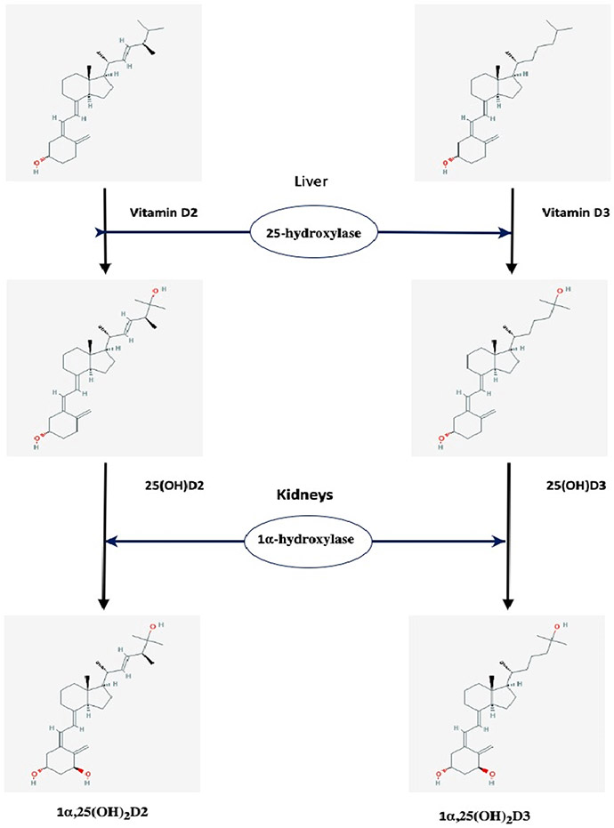
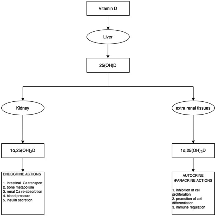
Vitamin D, an important hormone with a central role in calcium and phosphate homeostasis, is required for bone and muscle development as well as preservation of musculoskeletal function. The most abundant vitamin D metabolite is 25-hydroxyvitamin D [25(OH)D], which is currently considered the best marker to evaluate overall vitamin D status. 25(OH)D is therefore the most commonly measured metabolite in clinical practice. However, several other metabolites, although not broadly measured, are useful in certain clinical situations. Vitamin D and all its metabolites are circulating in blood bound to vitamin D binding protein, (VDBP). This highly polymorphic protein is not only the major transport protein which, along with albumin, binds over 99% of the circulating vitamin D metabolites, but also participates in the transport of the 25(OH)D into the cell via a megalin/cubilin complex. The accurate measurement of 25(OH)D has proved a difficult task. Although a reference method and standardization program are available for 25(OH)D, the other vitamin D metabolites still lack this. Interpretation of results, creation of clinical supplementation, and generation of therapeutic guidelines require not only accurate measurements of vitamin D metabolites, but also the accurate measurements of several other "molecules" related with bone metabolism. IFCC understood this priority and a committee has been established with the task to support and continue the standardization processes of vitamin D metabolites along with other bone-related biomarkers. In this review, we present the position of this IFCC Committee on Bone Metabolism on the latest developments concerning the measurement and standardization of vitamin D metabolites and its binding protein, as well as clinical indications for their measurement and interpretation of the results.
Keywords: 1α,25-dihydroxyvitamin D; 24,25-dihydroxyvitamin D; 25-hydroxyvitamin D; Immunoassays; Liquid chromatography; Mass spectrometry; Standardization; Vitamin D; Vitamin D Standardization Program; Vitamin D binding protein.
Copyright © 2021 Elsevier B.V. All rights reserved.
Conflict of interest statement
Declaration of Competing Interest
Konstantinos Makris, Harjit P Bhattoa, Karen Phinney, Christopher T Sempos, Candice Z. Ulmer, Samuel D Vasikaran, Hubert Vesper, and Annemieke C Heijboer declare no conflict of interest. Etienne Cavalier is a consultant for DiaSorin, IDS, Fujirebio, bioMérieux, Nittobo, and Menarini.
Figures
References
-
- Glendenning P, Inderjeeth CA, Screening for vitamin D deficiency: defining vitamin D deficiency, target thresholds of treatment and estimating the benefits of treatment, Pathology 44 (2) (2012) 160–165. - PubMed
-
- Souberbielle JC, Body JJ, Lappe JM, Plebani M, Shoenfeld Y, Wang TJ, Bischoff-Ferrari HA, Cavalier E, Ebeling PR, Fardellone P, Gandini S, Gruson D, Guerin AP, Heickendorff L, Hollis BW, Ish-Shalom S, Jean G, von Landenberg P, Largura A, Olsson T, Pierrot-Deseilligny C, Pilz S, Tincani A, Valcour A, Zittermann A, Vitamin D and musculoskeletal health, cardiovascular disease, autoimmunity and cancer: Recommendations for clinical practice, Autoimmun. Rev 9 (11) (2010) 709–715. - PubMed
Publication types
MeSH terms
Substances
Grants and funding
LinkOut - more resources
Full Text Sources
Other Literature Sources
Medical
