. 2021 May:119:102630.
doi: 10.1016/j.jaut.2021.102630. Epub 2021 Mar 10.

Mitochondrial N-formyl methionine peptides associate with disease activity as well as contribute to neutrophil activation in patients with rheumatoid arthritis

Affiliations

Mitochondrial N-formyl methionine peptides associate with disease activity as well as contribute to neutrophil activation in patients with rheumatoid arthritis

Bhargavi Duvvuri et al. J Autoimmun. 2021 May.

Abstract

Objectives: Literature suggests that neutrophils of patients with rheumatoid arthritis (RA) are primed to respond to N-formyl methionine group (formylated peptides). Animal models indicate that formylated peptides contribute to joint damage via neutrophil recruitment and inflammation in joints. Non-steroidal anti-inflammatory drugs are also known to inhibit formyl peptide-induced neutrophil activation. The predominant source of formylated peptides in sterile inflammatory conditions like RA is mitochondria, organelles with prokaryotic molecular signatures. However, there is no direct evidence of mitochondrial formyl peptides (mtNFPs) in the circulation of patients with RA and their potential role in neutrophil-mediated inflammation in RA, including their clinical significance.

Methods: Levels of mtNFPs (total fMet, MT-ND6) were analyzed using ELISA in plasma and serum obtained from patients in 3 cross-sectional RA cohorts (n = 275), a longitudinal inception cohort (n = 192) followed for a median of 8 years, and age/gender-matched healthy controls (total n = 134). Neutrophil activation assays were done in the absence or presence of formyl peptide receptor 1 (FPR1) inhibitor cyclosporine H.

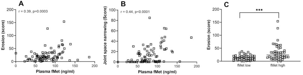
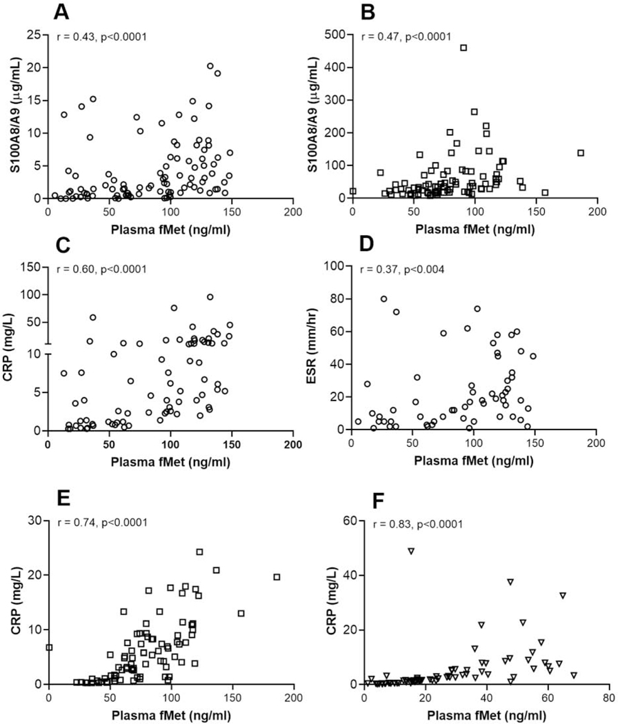
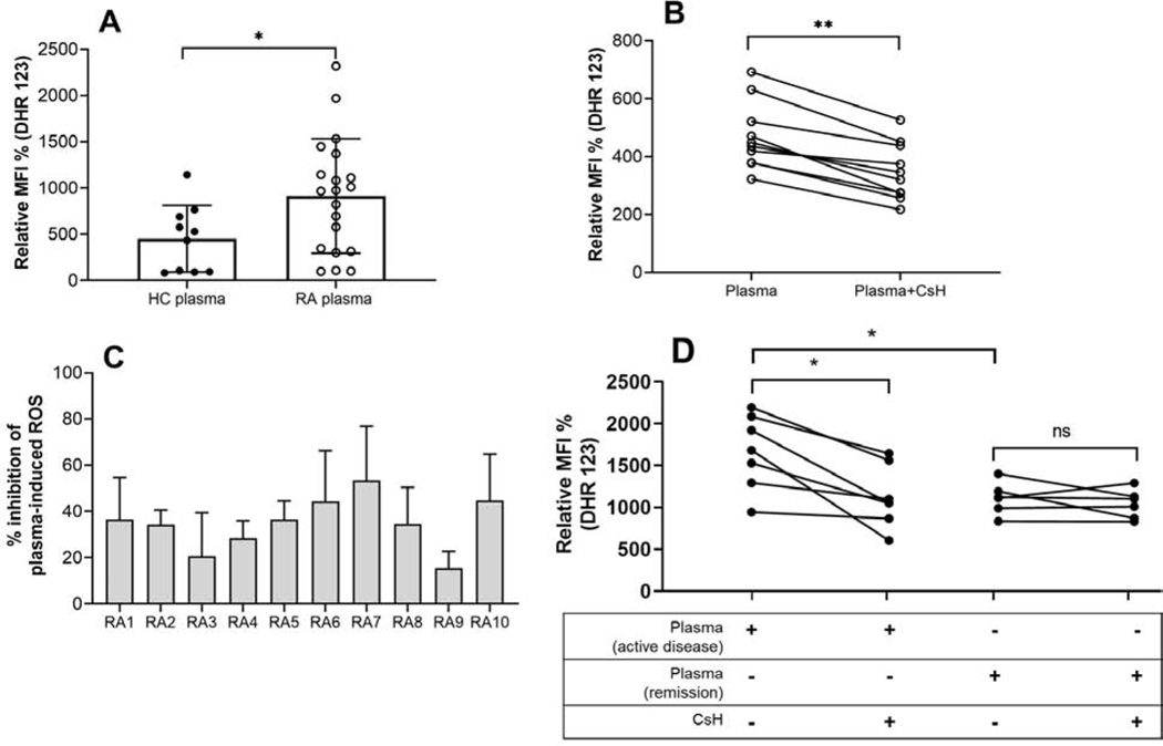
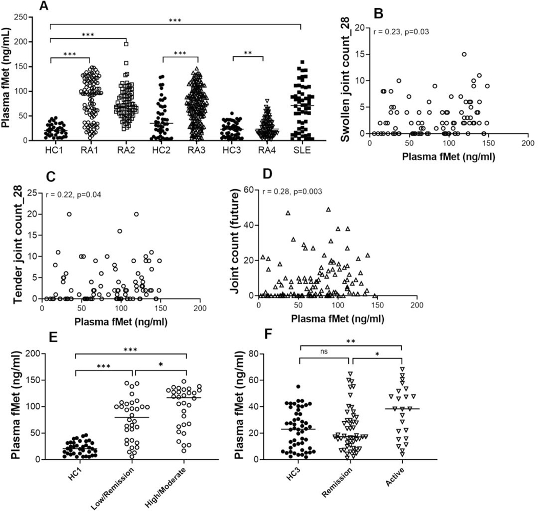
Results: Elevated levels of total fMet were observed in the circulation of patients with RA as compared to healthy controls (p < 0.0001) associating with disease activity and could distinguish patients with the active disease from patients with inactive disease or patients in remission. Baseline levels of total fMet correlated with current and future joint involvement, respectively and predicted the development of rheumatoid nodules (OR = 1.2, p = 0.04). Further, total fMet levels improved the prognostic ability of ACPA in predicting erosive disease (OR of 7.9, p = 0.001). Total fMet levels correlated with markers of inflammation and neutrophil activation. Circulating mtNFPs induced neutrophil activation in vitro through FPR1-dependent mechanisms.

Conclusions: Circulating mtNFPs could be novel biomarkers of disease monitoring and prognosis for RA and in investigating neutrophil-mediated inflammation in RA. We propose, FPR1 as a novel therapeutic target for RA.

Keywords: Clinical biomarkers; Formyl peptide receptor 1; Mitochondria; Neutrophils; Rheumatoid arthritis.


Figures

Figure 5:
Figure 5:. MtNFPs (total fMet) circulating in RA plasma can induce ROS release from neutrophils in FPR1-dependent manner.
(A) Neutrophil ROS induced by RA plasma compared to HC plasma. (B) Inhibition of RA plasma-induced neutrophil ROS by CsH. (C) Percent inhibition of RA plasma-induced neutrophil ROS. (D) Comparison of ROS-induction potential of plasma from patients with active disease and patients in remission from neutrophils with and without pretreatment with FPR1 inhibitor, CsH. Healthy neutrophils plated at 3 × 10⁵ cells/well were incubated with a selective inhibitor of FPR1, CsH (5 μM) for 30 min prior to the addition of plasma (1:100 dilution) for an additional 60 min. DHR 123 (0.5 μM), was added during last 30 min of incubation and ROS was analyzed by flow cytometry. Relative MFI % was calculated as ROS induced by stimuli divided by media control x 100; % inhibition was calculated as ROS induced by plasma only – ROS induced by plasma pretreated with CsH condition divided by ROS induced by plasma only x 100. Statistical analyses were done using Mann-Whitney U test and Wilcoxon test for paired analyses with * < 0.05, and ** p<0.01. Samples belong to RA1, white circle ○.
Figure 1:
Figure 1:. Levels of mtNFPs are elevated in patients with RA and associate with disease activity.
Total (fMet) and MT-ND6 specific levels of NFPs were analyzed by ELISA. A, Total fMet levels were analyzed in 4 cohorts of RA patients, a SLE cohort and 3 cohorts of healthy controls (HC). Plasma samples were analyzed in cohorts RA1, RA2, RA4, SLE, HC1 and HC3. Serum samples were analyzed in cohorts RA3 and HC2. For RA4, only fMet levels of patients with low to high DAS28-CRP (> 2.6) were considered, but not levels in patients in remission (<2.6 DAS28-CRP). Correlation analysis between levels of total fMet and swollen joints (B), tender joints (C) of RA1 and future joint count (D) in the inception cohort, RA3. Healthy controls and RA patients were analyzed based on total fMet levels. Patients were further analyzed based on CDAI in RA1 (E) and DAS28-CRP in RA4 (F). Statistical analyses were done using Mann-Whitney U test, and Spearman correlation test with * P < 0.05, ** p<0.01, *** p<0.0001, and ns, non-significant.
Figure 2:
Figure 2:. Levels of mtNFPs (total fMet) in RA patients cluster with markers of disease activity and inflammation.
Shown in A and B are heat maps showing hierarchical clustering of various disease activity, diagnostic and inflammatory markers with levels of mtNFPs in cohorts RA1 (A) and RA4 (B). Rows and columns represent markers and patients, respectively. Hierarchical clustering was performed using the R v4.0.2 pheatmap v1.0.12
Figure 3:
Figure 3:. MtNFPs (total fMet) circulating in RA plasma associate with joint damage.
Correlation analysis between levels of total fMet and erosion score (A) joint space narrowing (B) and (C) comparison of erosion scores between patients stratified based on fMet levels as fMet low or fMet high (fMet levels above 95th percentile of HC are considered as fMet high) for follow-up cohort, RA2 white square □. Statistical analyses were done using Mann-Whitney U test, and Spearman correlation test with *** p<0.001.
Figure 4:
Figure 4:. Levels of mtNFPs (total fMet) in RA patients associate with neutrophil activation markers and clinical measures inflammation.
Total fMet and S100A8/A9 levels were analyzed by ELISA. Shown in figures A, C and D, figures B and E and figure F are data from cohorts: RA1 (white circle ○), RA2 (white square □), and RA4 (white down-pointing triangle ∇), respectively. Statistical analyses were done using Spearman correlation test.

Similar articles

Cited by

References

    1. McInnes IB, Schett G. The pathogenesis of rheumatoid arthritis. N Engl J Med, 2011;365:2205–19. - PubMed
    1. Wipke BT, Allen PM. Essential role of neutrophils in the initiation and progression of a murine model of rheumatoid arthritis. J Immunol, 2001;167:1601–8. - PubMed
    1. Tanaka D, Kagari T, Doi H, Shimozato T. Essential role of neutrophils in anti-type II collagen antibody and lipopolysaccharide-induced arthritis. Immunology, 2006;119:195–202. - PMC - PubMed
    1. Wright HL, Chikura B, Bucknall RC, Moots RJ, Edwards SW. Changes in expression of membrane TNF, NF-{kappa}B activation and neutrophil apoptosis during active and resolved inflammation. Ann Rheum Dis, 2011;70:537–43. - PubMed
    1. Mantovani A, Cassatella MA, Costantini C, Jaillon S. Neutrophils in the activation and regulation of innate and adaptive immunity. Nat Rev Immunol, 2011;11:519–31. - PubMed

Publication types

MeSH terms