

Common and distinct brain networks of autoscopic phenomena
- PMID: 33714069
- PMCID: PMC7970131
- DOI: 10.1016/j.nicl.2021.102612
Common and distinct brain networks of autoscopic phenomena
Abstract
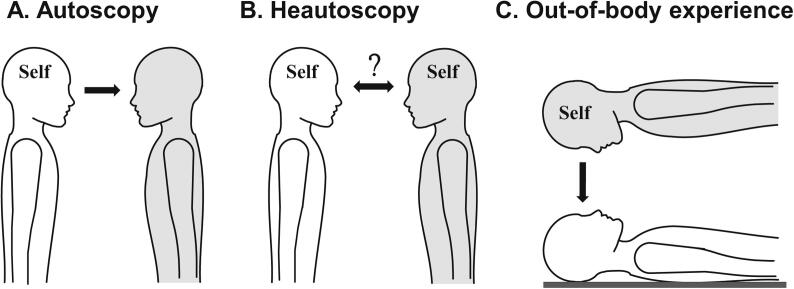
Objective: Autoscopic phenomena (AP) are illusory own body reduplications characterized by the visual perception of a second own body in extrapersonal space, and include three main forms: autoscopic hallucination (AH), heautoscopy (HAS) and out-of-body-experience (OBE). Past research showed that lesions were heterogeneously distributed and affected many different brain regions within and across patients, while small case series suggested that AP lesions converge in temporo-parietal and parieto-occipital cortex. As only few studies investigated each form of AP separately, it remains unknown whether the three AP are characterized by common and distinct brain mechanisms.
Methods: Here, we applied lesion network analysis in 26 neurological AP patients and determined their common and distinct functional connectivity patterns.
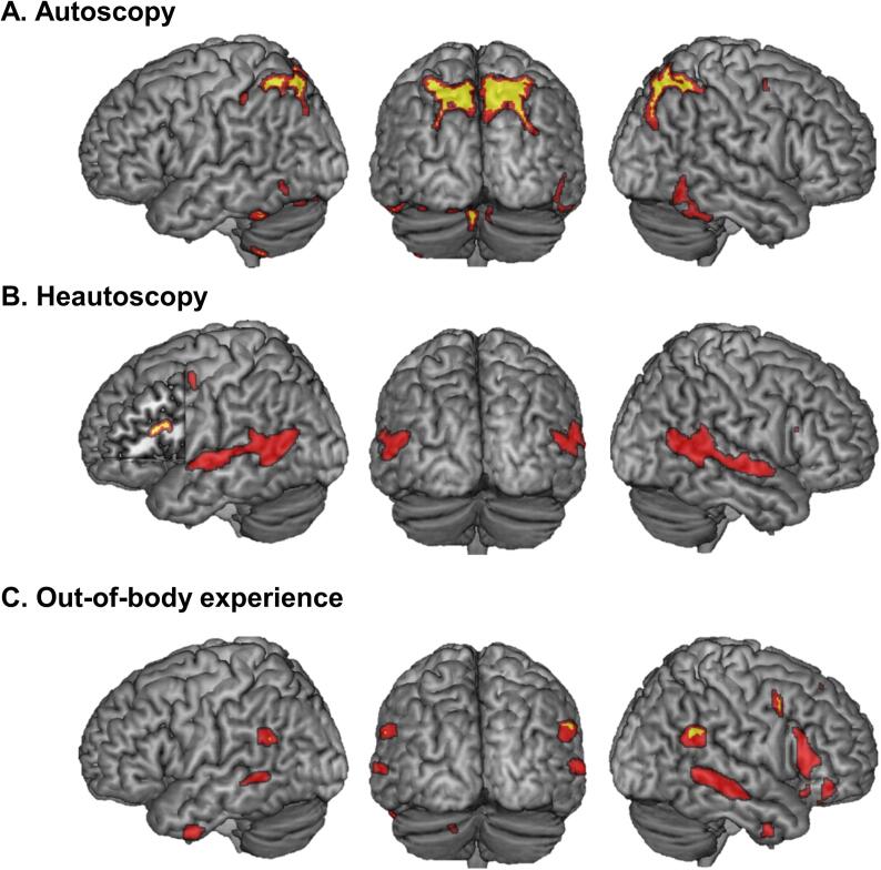
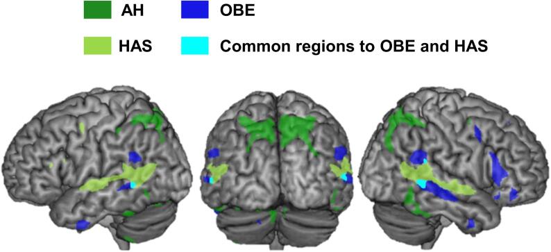
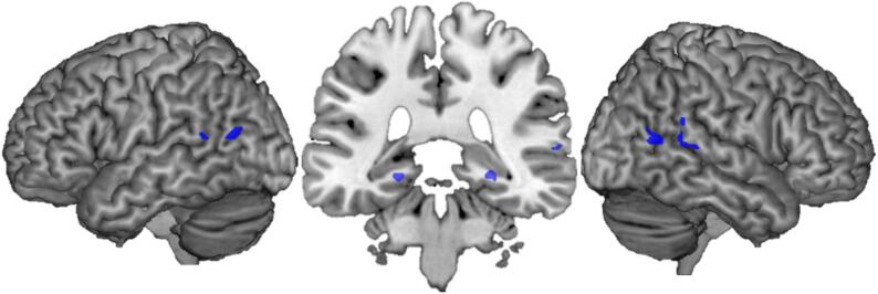
Results: We report that all localize to a single common brain network at the bilateral temporo-parietal junction, further associated with specific patterns of functional connectivity, defining each type of AP. OBE resulted from a brain network connected to bilateral angular gyrus, right precuneus, and right inferior frontal gyrus, differing from AH with a brain network connected to bilateral precuneus, inferior temporal gyrus, and cerebellum. HAS resulted from a brain network connected to left inferior frontal gyrus, left insula and left parahippocampus.
Conclusion: The present data identify the temporo-parietal junction as the common core region for AP and show that each form of AP recruits additional specific networks, associated with different sensorimotor and self-related sub-networks.
Keywords: Bodily self-consciousness; Lesion network mapping; Multisensory processing; Temporo-parietal junction.
Copyright © 2021 The Authors. Published by Elsevier Inc. All rights reserved.
Conflict of interest statement
The authors declare that they have no known competing financial interests or personal relationships that could have appeared to influence the work reported in this paper.
Figures
References
-
- Allen P., Larøi F., McGuire P.K., Aleman A. The hallucinating brain: A review of structural and functional neuroimaging studies of hallucinations. Neurosci. Biobehav. Rev. 2008;32:175–191. - PubMed
-
- Allison T., Ginter H., McCarthy G. Face recognition in human extrastriate cortex. J. Neurophysiol. 1994;71:821–825. - PubMed
-
- Anzellotti F., Onofrj V., Maruotti V. Autoscopic phenomena: case report and review of literature. Behav Brain Funct [online serial]. 2011;7:2. http://www.biomedcentral.com/content/pdf/1744-9081-7-2.pdf%5Cnhttp://www... Accessed at: - PMC - PubMed
Publication types
MeSH terms
LinkOut - more resources
Full Text Sources
Other Literature Sources
Research Materials
Miscellaneous
