. 2021 May;76(5):392.e1-392.e9.
doi: 10.1016/j.crad.2021.02.009. Epub 2021 Feb 25.

Qualitative and quantitative DECT pulmonary angiography in COVID-19 pneumonia and pulmonary embolism

Affiliations

Qualitative and quantitative DECT pulmonary angiography in COVID-19 pneumonia and pulmonary embolism

C D Arru et al. Clin Radiol. 2021 May.

Abstract

Aim: To assess differences in qualitative and quantitative parameters of pulmonary perfusion from dual-energy computed tomography (CT) pulmonary angiography (DECT-PA) in patients with COVID-19 pneumonia with and without pulmonary embolism (PE).

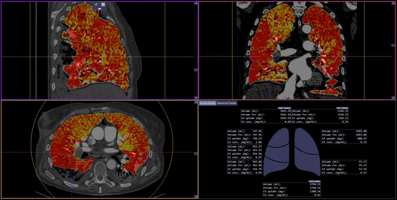
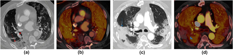
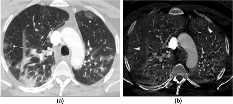
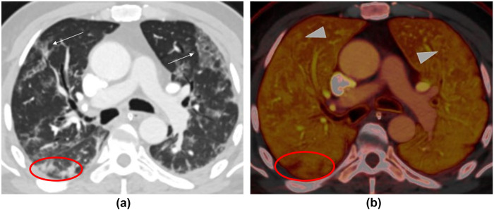
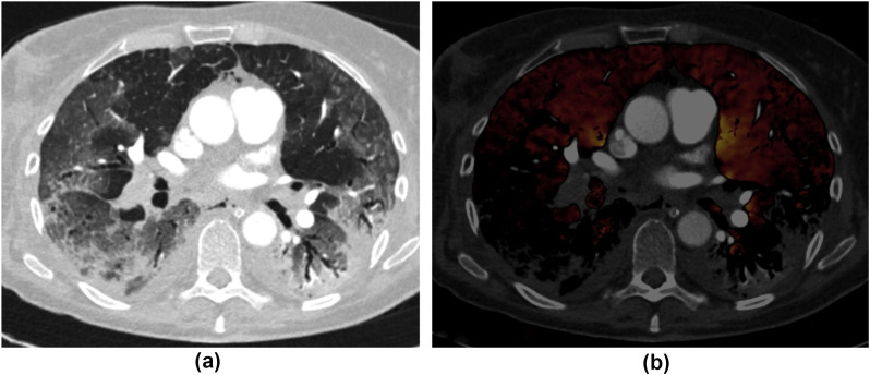
Materials and methods: This retrospective institutional review board-approved study included 74 patients (mean age 61±18 years, male:female 34:40) with COVID-19 pneumonia in two countries (one with 68 patients, and the other with six patients) who underwent DECT-PA on either dual-source (DS) or single-source (SS) multidetector CT machines. Images from DS-DECT-PA were processed to obtain virtual mono-energetic 40 keV (Mono40), material decomposition iodine (MDI) images and quantitative perfusion statistics (QPS). Two thoracic radiologists determined CT severity scores based on type and extent of pulmonary opacities, assessed presence of PE, and pulmonary parenchymal perfusion on MDI images. The QPS were calculated from the CT Lung Isolation prototype (Siemens). The correlated clinical outcomes included duration of hospital stay, intubation, SpO2 and death. The significance of association was determined by receiver operating characteristics and analysis of variance.

Results: One-fifth (20.2%, 15/74 patients) had pulmonary arterial filling defects; most filling defects were occlusive (28/44) located in the segmental and sub-segmental arteries. The parenchymal opacities were more extensive and denser (CT severity score 24±4) in patients with arterial filling defects than without filling defects (20±8; p=0.028). Ground-glass opacities demonstrated increased iodine distribution; mixed and consolidative opacities had reduced iodine on DS-DECT-PA but increased or heterogeneous iodine content on SS-DECT-PA. QPS were significantly lower in patients with low SpO2 (p=0.003), intubation (p=0.006), and pulmonary arterial filling defects (p=0.007).

Conclusion: DECT-PA QPS correlated with clinical outcomes in COVID-19 patients.


Figures

Figure 1
Figure 1
QPS software in action. The software automatically generates MDI images in the transverse, coronal and sagittal planes as well as a diagrammatic (bottom right image) and tabular representation (not shown) of QPS features for the whole lung, right lung, left lung, and each lung lobe.
Figure 2
Figure 2
(a,c) Transverse virtual mono-energetic (Mono 40 keV) and (b,d) material decomposition iodine (MDI) images belonging to two patients scanned with DS-DECT-PA. (a) Mono 40 keV image of a 67-year-old COVID-19 infected man demonstrates a mixed ground-glass and consolidative opacity in the right lower lobe (red arrow) and pulmonary embolus in the segmental right lower lobe pulmonary arteries; (b) the MDI image demonstrates decreased lung perfusion in the region of the mixed opacity consistent with a pulmonary infarction. (c) Mono 40 keV image of an 80-year-old man has multifocal consolidations in bilateral upper and right lower lobes, which correspond to regions of perfusions defects on MDI image (d). Both patients had RT-PCR positive COVID-19 pneumonia.
Figure 3
Figure 3
(a) Transverse virtual mono-energetic (Mono 40 keV) and (b) material decomposition iodine (MDI) images of a 48-year-old COVID-19 infected man with SS-DECT-PA. Mono 40 keV image demonstrate ground-glass opacities in bilateral upper lobes (along arrows), which correspond to regions of increased perfusion (iodine related enhancement) on MDI images. The consolidative opacity in the right lower lobe demonstrates variable perfusion pattern.
Figure 4
Figure 4
(a) Transverse virtual mono-energetic (Mono 40 keV) and (b) material decomposition iodine (MDI) images of a 54-year-old COVID-19 infected man who underwent DS-DECT-PA. Mono 40 keV images demonstrate ground-glass opacities in the bilateral upper lobes (along arrows), which correspond to regions of increased perfusion (arrowheads) on MDI images. The nodular foci of consolidation in the right lower lobe demonstrate decreased perfusion compared to the adjacent normal lung (within the red ovals).
Figure 5
Figure 5
(a) Transverse chest CT image in lung window of a 45-year-old woman with COVID-19 pneumonia demonstrates mixed consolidation and ground glass in the bilateral lower lobes along with multifocal ground-glass opacities in the upper lobes. (b) At the same anatomical level, the transverse MDI image (a fused image of lung perfusion overlaid on conventional CT image in a soft-tissue window) depicts large pulmonary emboli in the right lower lobar and segmental arteries. The decreased perfusion in the right lower lobe is attributed to a perfusion defect from occlusive PE. The lack of perfusion in the left lower lobe without corresponding PE is artefactual as regions with consolidation are excluded from evaluation.

Similar articles

Cited by

References

    1. McGonagle D., O’Donnell J., Sharif K. Immune mechanisms of pulmonary intravascular coagulopathy in COVID-19 pneumonia. Lancet Rheumatol. 2020 doi: 10.1016/S2665-9913(20)30121-1. - DOI - PMC - PubMed
    1. Varga S., Flammer A., Steiger P. Endothelial cell infection and endothelitis in COVID-19. Lancet. May 2020;10234:1417–1418. - PMC - PubMed
    1. Lodigiani C., Iapichino G., Carenzo L. Venous and arterial thromboembolic complications in COVID-19 patients admitted to an academic hospital in Milan, Italy. Thromb Res. 2020;191:9–14. - PMC - PubMed
    1. Mahammedi A., Saba L., Vagal A. Imaging in neurological disease of hospitalized COVID-19 patients: an Italian multicenter retrospective observational study. Radiology. May 2020 doi: 10.1148/radiol.2020201933. - DOI - PMC - PubMed
    1. Oxley T.J., Mocco J., Majidi S. Large-vessel stroke as a presenting feature of Covid-19 in the young. N Engl J Med. 2020;382(20):e60. - PMC - PubMed

MeSH terms