Review
. 2021 Apr;96(4):989-1005.
doi: 10.1016/j.mayocp.2021.01.009. Epub 2021 Jan 21.

Pernio (Chilblains), SARS-CoV-2, and COVID Toes Unified Through Cutaneous and Systemic Mechanisms

Affiliations
Review

Pernio (Chilblains), SARS-CoV-2, and COVID Toes Unified Through Cutaneous and Systemic Mechanisms

Mark A Cappel et al. Mayo Clin Proc. 2021 Apr.

Abstract

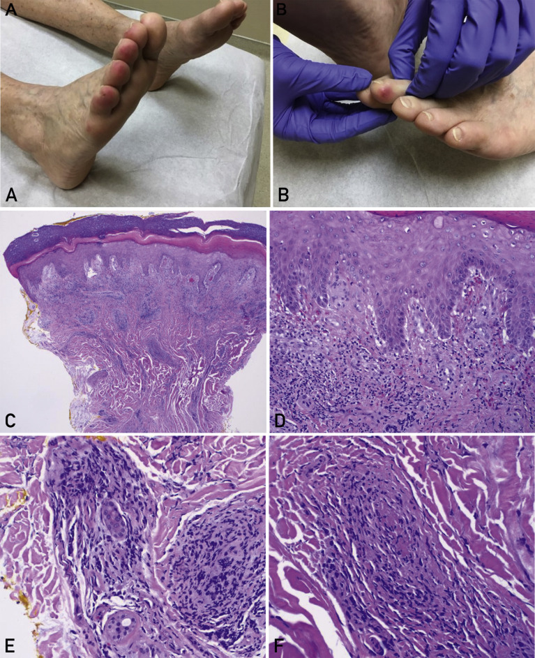
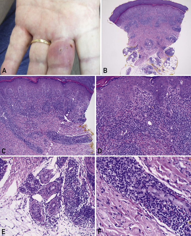
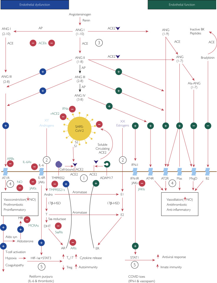
Pernio or chilblains is characterized by erythema and swelling at acral sites (eg, toes and fingers), typically triggered by cold exposure. Clinical and histopathologic features of pernio are well described, but the pathogenesis is not entirely understood; vasospasm and a type I interferon (IFN-I) immune response are likely involved. During the coronavirus disease 2019 (COVID-19) pandemic, dermatologists have observed an increase in pernio-like acral eruptions. Direct causality of pernio due to COVID-19 has not been established in many cases because of inconsistent testing methods (often negative results) for severe acute respiratory syndrome coronavirus 2 (SARS-CoV-2). However, a form of COVID-19‒associated pernio (also called COVID toes) is probable because of increased occurrence, frequently in young patients with no cold exposure or a history of pernio, and reports of skin biopsies with positive SARS-CoV-2 immunohistochemistry. PubMed was searched between January 1, 2020, and December 31, 2020 for publications using the following keywords: pernio, chilblain, and acral COVID-19. On the basis of our review of the published literature, we speculate that several unifying cutaneous and systemic mechanisms may explain COVID-19‒associated pernio: (1) SARS-CoV-2 cell infection occurs through the cellular receptor angiotensin-converting enzyme 2 mediated by transmembrane protease serine 2, subsequently affecting the renin-angiotensin-aldosterone system with an increase in the vasoconstricting, pro-inflammatory, and prothrombotic angiotensin II pathway. (2) Severe acute respiratory syndrome coronavirus 2 cell infection triggers an immune response with robust IFN-I release in patients predisposed to COVID-19‒associated pernio. (3) Age and sex discrepancies correlated with COVID-19 severity and manifestations, including pernio as a sign of mild disease, are likely explained by age-related immune and vascular differences influenced by sex hormones and genetics, which affect susceptibility to viral cellular infection, the renin-angiotensin-aldosterone system balance, and the IFN-I response.


Figures

Figure 1
Figure 1
Pernio in a female patient. A woman, who was in her 70s, was evaluated in Florida in February 2020. She had a history of pernio related to rheumatoid arthritis, with chronic waxing and waning tender lesions on her toes, exacerbated by wearing sandals in an air-conditioned indoor environment. Coincidentally, during the coronavirus disease 2019 (COVID-19) pandemic, she later received an unrelated diagnosis of COVID-19. She did not require hospitalization and recovered as an outpatient. Interestingly, she reported no clinically significant worsening of pernio during this viral respiratory illness, possibly because her rheumatoid arthritis was treated with tofacitinib, a Janus kinase inhibitor, which may have inhibited the effect of signal transducer and activator of transcription 1‒dependent type I interferons thought to play a role in the pathophysiology of pernio and COVID-19. A and B, Clinical photographs of the right foot (panel A) and left foot (panel B) illustrate erythematous edematous plaques affecting the distal toes. Courtesy of Ines Kevric O’Shaughnessy, MD, First Coast Dermatology Associates, Jacksonville Beach, FL; used with permission. C-F, Histopathologic sections of the patient’s punch biopsy specimen (hematoxylin-eosin) exhibit a superficial and deep dermal lymphocytic inflammatory infiltrate (panel C; original magnification, ×40); lichenoid interface dermatitis along the dermal-epidermal junction with basal vacuolar changes (panel D; original magnification, ×200); perivascular and perieccrine inflammation (panel E; original magnification, ×200); and focal lymphocytic vasculitis with fibrin thrombi involving a small dermal vessel (panel F; original magnification, ×400).
Figure 2
Figure 2
Pernio in a male patient. A man who was in his 70s was evaluated in Florida in February 2020. He reported a few intermittent flares of an inflamed lesion, which affected only the long finger of the left hand. He had no history of pernio, cold exposure, autoimmune disease, travel history, or testing for coronavirus disease 2019 (COVID-19). He presented the week before the first positive case of COVID-19 was confirmed in Florida, so an association with COVID-19 is unlikely, unless unrecognized community spread had occurred. A, Clinical photograph of the left hand illustrates an erythematous edematous plaque with focal vesiculation affecting the long finger of the left hand. Courtesy of James B. Connors, MD, BayCare Medical Group, Sun Coast Medical Clinic Dermatology, Saint Petersburg, FL; used with permission. B-F, Histopathologic sections of the patient’s punch biopsy specimen (hematoxylin-eosin) illustrate a superficial and deep dermal lymphocytic inflammatory infiltrate (panel B; original magnification, ×40); brisk perivascular inflammation in the superficial to mid dermis (panel C; original magnification, ×100); lichenoid interface dermatitis along the dermal-epidermal junction with basal vacuolar changes (panel D; original magnification, ×200); perieccrine lymphocytic inflammation at the junction of the deep reticular dermis and the subcutaneous adipose tissue (panel E; original magnification, ×200); and focal lymphocytic vasculitis involving a small dermal vessel, with endothelial swelling and extravasation of red blood cells into the surrounding dermis (panel F; original magnification, ×400).
Figure 3
Figure 3
Interplay of severe acute respiratory syndrome coronavirus 2 (SARS-CoV-2), angiotensin-converting enzyme (ACE) 2 (ACE2), the renin-angiotensin-aldosterone system (RAAS), sex hormones, and the immune response: a potential mechanism of coronavirus disease (COVID) toes. Used with permission of M.A. Cappel, MD. Circled numbers indicate steps in the mechanism. Step 1: The cellular receptor ACE2 is critically important in SARS-CoV-2 infection., In addition, transmembrane protease serine 2 (TMPRSS2) is essential because by cleaving cell-bound ACE2 and SARS-CoV-2 spike protein subunit 1 (S1) from spike protein subunit 2 (S2), it facilitates viral cellular entry.,,Step 2: Androgens and estrogens have generally opposing downstream effects on ACE2 processing, providing an explanation for more severe coronavirus disease 2019 (COVID-19) in male patients. TMPRSS2 activity increases with androgen sensitization through dihydrotestosterone activation of the androgen receptor (AR). On the contrary, estrogens increase the expression of a disintegrin and metalloproteinase 17 (ADAM17), which competes for processing of ACE2 and releases a circulating form of active ACE2., Therefore, increased ADAM17 activity may be protective in female patients, resulting in an increased proportion of circulating ACE2 that binds any circulating SARS-CoV-2 and prevents further cell infection. Step 3: When cells are infected by SARS-CoV-2, the resulting virus-receptor internalization results in the decreased cell expression of ACE2 and a relative deficiency of ACE2. In the RAAS, a delicate balance exists between ACE and ACE2. ACE converts angiotensin (ANG) I (ANGI) to ANGII and, with aminopeptidase, to ANGIII, both of which contribute to endothelial dysfunction through binding the angiotensin type 1 receptor (AT1R). On the contrary, ACE2 converts ANGI to ANG1-9 and ANGII to ANG1-7, both of which promote healthy endothelial function through binding the AT2R. At baseline, because of sex hormone differences, the ACE–ANGII–AT1R pathway is favored in male patients whereas the ACE2‒ANG1-7‒AT2R pathway is favored in female patients.Step 4: These RAAS predilections may account for increased endothelial dysfunction in male patients compared with female patients; AT1R stimulation decreases nitric oxide (NO) and is vasoconstricting, prothrombotic, and pro-inflammatory; AT2R stimulation increases NO and is vasodilatory, antithrombotic, and anti-inflammatory. Angiotensin II–AT1R activation also potentiates endothelial dysfunction by stimulating aldosterone synthesis and subsequent mineralocorticoid receptor activation, with similar vasculopathic and pro-inflammatory effects.Step 5: Aldosterone stimulates mineralocorticoid receptors on dendritic cells, and ANGII stimulates AT1Rs on T cells and on dendritic cells; all these actions promote T-cell activation and proliferation. This leads to activation of interleukin 6 family (IL-6) cytokine receptors (IL-6Rs), which signal through the Janus kinase (JAK)‒signal transducer and activator of transcription (STAT) 3 pathway, and activation of the type I interferon (IFN-I) receptor (IFN-IR), which signals through the JAK-STAT1 pathway., Tissue hypoxia from the related AT1R-induced endothelial dysfunction may also contribute through hypoxia-inducible factor 1α subunit (HIF-1α) in conjunction with STAT3 to increase pro-inflammatory helper T cell 17s (TH17s) and decrease anti-inflammatory regulatory T cells (Tregs). Owing to sex hormone and genetic differences, female patients have a more robust IFN-I‒STAT1 response than do male patients., Younger individuals have a stronger IFN-I response, which favors the development of COVID toes; in older individuals the IL-6 pathway may predominate, which is associated with more severe COVID-19. 5αRI, 5α-reductase inhibitor; 17β-HSD, 17β-hydroxysteroid dehydrogenase; ACEI, angiotensin-converting enzyme inhibitor; Ala, alamandine; Aldo syn, aldosterone synthase; Andro, androstenedione; AP, aminopeptidase; ARB, angiotensin receptor blocker; ARI, androgen receptor inhibitor; B2, bradykinin receptor B2; BK, bradykinin; DHT, dihydrotestosterone; E1, estrone; E2, estradiol; ER, estrogen receptor; IFN, interferon; IL-6A, IL-6 antagonist; JAKI, Janus kinase inhibitor; Mas, G protein‒coupled receptor Mas receptor; MCRA, mineralocorticoid receptor antagonist; MR, mineralocorticoid receptor; MrgD, Mas-related G protein‒coupled receptor member D; rACE2, recombinant angiotensin-converting enzyme 2; T, testosterone; TMPRSS2I, transmembrane protease serine 2 inhibitor; XX, 2 X chromosomes (genetic female); XY, 1 X chromosome and 1 Y chromosome (genetic male).

Similar articles

Cited by

References

    1. Florida COVID-19 response Florida Health website. https://floridahealthcovid19.gov/#latest-stats
    1. Pernio Wiktionary website. https://en.wiktionary.org/wiki/pernio
    1. Cappel J.A., Wetter D.A. Clinical characteristics, etiologic associations, laboratory findings, treatment, and proposal of diagnostic criteria of pernio (chilblains) in a series of 104 patients at Mayo Clinic, 2000 to 2011. Mayo Clin Proc. 2014;89(2):207–215. - PubMed
    1. Su W.P., Perniciaro C., Rogers R.S., III, White J.W., Jr. Chilblain lupus erythematosus (lupus pernio): clinical review of the Mayo Clinic experience and proposal of diagnostic criteria. Cutis. 1994;54(6):395–399. - PubMed
    1. Neville E., Mills R.G., Jash D.K., Mackinnon D.M., Carstairs L.S., James D.G. Sarcoidosis of the upper respiratory tract and its association with lupus pernio. Thorax. 1976;31(6):660–664. - PMC - PubMed