. 2021 Mar 10;13(6):8628-8642.
doi: 10.18632/aging.202677. Epub 2021 Mar 10.

Activation of ATF4 triggers trabecular meshwork cell dysfunction and apoptosis in POAG

Affiliations

Activation of ATF4 triggers trabecular meshwork cell dysfunction and apoptosis in POAG

Ying Ying et al. Aging (Albany NY). .

Abstract

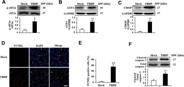
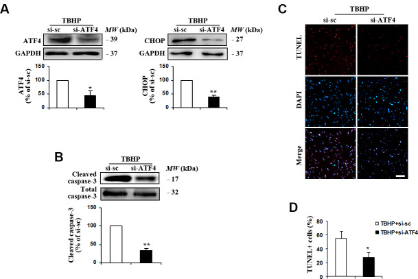
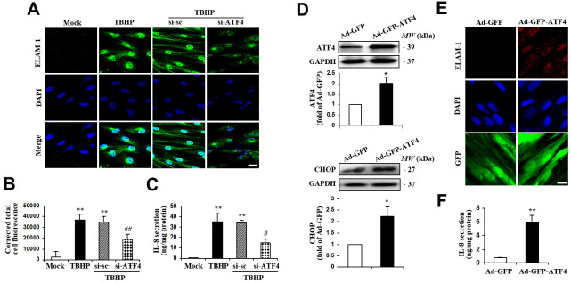
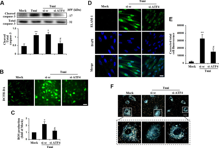
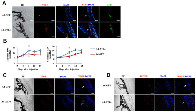
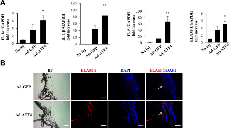
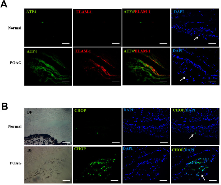
Primary open angle glaucoma (POAG) is the leading cause of irreversible blindness. Dysfunction of the trabecular meshwork (TM), resulting in decreased outflow of aqueous humor and increased intraocular pressure (IOP), plays an important role in the pathogenesis of POAG. However, the underlying mechanisms still remain unclear. In this study, we demonstrated that the eIF2-α/ATF4/CHOP branch of unfolded protein response (UPR) was activated in human trabecular meshwork cells (HTMCs) upon tert-butyl hydroperoxide (TBHP) exposure. Inhibition of ATF4 ameliorated TBHP-induced apoptosis and inflammatory cytokine production, while ectopic expression of ATF4 increased the expression of endothelial leukocyte adhesion molecule (ELAM)-1 and IL-8 in HTMCs. Furthermore, we found that ATF4 inhibition reduced tunicamycin-induced caspase-3 activation, ROS production, ELAM-1 expression, and HTMCs phagocytosis impairment. By an in vivo study in mice, we showed that overexpression of ATF4 in the TM induced C/EBP homologous protein (CHOP) expression and TM cells apoptosis, contributing to inflammatory cytokine production, and probably IOP elevation. More importantly, upregulation of ATF4 and CHOP, and colocalization of ATF4 with ELAM-1 were found in the TM of POAG patients. These results suggest that ATF4 is a critical mediator of oxidative stress and ER stress-induced TM cell dysfunction and apoptosis in POAG.

Keywords: activating transcription factor 4; endoplasmic reticulum stress; oxidative stress; primary open angle glaucoma; trabecular meshwork.


Conflict of interest statement

CONFLICTS OF INTEREST: The authors declare that they have no conflicts of interest.

Figures

Figure 1
Figure 1
TBHP activated the expression of transcription factor ATF4 and induced apoptosis in HTMCs. HTMCs were exposed to media in the absence (Mock) or presence of 50 μM of TBHP for 12 h. (AC) Western blot analysis for phospho-eIF2α, total eIF2α, ATF4, CHOP and GAPDH. Intensities were quantified and normalized against the level of total proteins (eIF2α) or GAPDH (mean ± SEM, n = 3). (D) Representative images of immunostaining for apoptotic (TUNEL-positive, red) HTMCs. Nuclei were labeled with DAPI (blue). Scale bar represents 100 μm. (E) Quantification of apoptotic nuclei by Image-Pro Plus software. Values are expressed as the percentage of TUNEL-positive cells to total (DAPI-positive) cells (mean ± SEM, n = 3). (F) Western blot analysis for cleaved caspase-3. Intensities were quantified and normalized against the level of total caspase-3 (mean ± SEM, n = 3). *P<0.05 and **P <0.01 vs. Mock.
Figure 2
Figure 2
Suppression of ATF4 expression by siRNA transfection inhibited TBHP-induced apoptosis of HTMCs. HTMCs were transfected with siRNA specific to ATF4 (si-ATF4) or to scramble sequences (si-sc) for 48 h, after which they were exposed to 50 μM of TBHP for additional 12 h. (A) The suppression efficiency of ATF4 using si-RNA transfection and the expression of its downstream target CHOP were determined by Western blot analysis (mean ± SEM, n = 3). (B) Levels of cleaved caspase-3 were examined by Western blot. Intensities of protein expression were quantified, normalized against the level of total caspase-3 and expressed as relative changes to protein abundance in cells transfected with scramble control (si-sc) (mean ± SEM, n = 3). (C) Apoptotic cells were examined by TUNEL staining (red, TUNEL; blue, DAPI; scale bar, 100 μm). (D) Numbers of apoptotic cells were quantified and expressed as the percentage of TUNEL-positive to DAPI-positive cells (mean ± SEM, n = 3). *P<0.05 and **P<0.01 vs. TBHP+si-sc. TBHP+si-sc, exposure of HTMCs transfected with si-sc to TBHP. TBHP+si-ATF4, exposure of HTMCs transfected with si-ATF4 to TBHP.
Figure 3
Figure 3
ATF4 is an important mediator of ELAM-1 expression and IL-8 secretion in HTMCs. (AC) HTMCs were exposed to media in the absence (Mock) or presence of 50 μM of TBHP for 12 h. Alternatively, cells were transfected with si-ATF4 or si-sc for 48 h, followed by incubation with 50 μM of TBHP for additional 12 h. (A) Representative images of subcellular expression of ELAM-1 as examined by indirect immunofluorescence (green, ELAM-1. blue, DAPI. scale bar, 40 μm). (B) Levels of cellular immunofluorescence of ELAM-1 were quantified and expressed as corrected total cell fluorescence (mean ± SEM, n = 3). (C) IL-8 secretion was assayed in culture supernatant of HTMCs using an IL-8 ELISA kit. Values were normalized for total protein at the respective treatment (mean ± SEM, n = 3). **P <0.01 vs. Mock, #P <0.05 and ##P <0.01 vs. TBHP+si-sc. TBHP+si-sc, exposure of cells transfected with si-sc to TBHP. TBHP+si-ATF4, exposure of cells transfected with si-ATF4 to TBHP. (DF) A recombinant adenovirus coding ATF4 (Ad-GFP-ATF4) was constructed to express GFP protein as a marker for the identification of infected cells. Cultured HTMCs were infected with Ad-GFP-ATF4 or empty vector (Ad-GFP) for 72 h. (D) Western blot analysis for ATF4 and CHOP. Intensities of protein expression were quantified, normalized against the level of GAPDH and expressed as relative changes to Ad-GFP (mean ± SEM, n = 3). (E) Subcellular expression of ELAM-1 was detected by immunofluorescent staining (red, ELAM-1. blue, DAPI. green, GFP. scale bar, 20 μm). (F) IL-8 secretion was assayed using ELISA. Values were normalized for total protein at the respective treatment (mean ± SEM, n = 3). *P <0.05, **P <0.01 vs. Ad-GFP.
Figure 4
Figure 4
Suppression of ATF4 prevented tunicamycin-induced apoptosis and ROS generation, and rescued the phagocytotic activity of HTMC. HTMCs were exposed to media in the absence (Mock) or presence of 100 ng/ml of tunicamycin (Tuni) for 24 h. Alternatively, cells were transfected with si-ATF4 or si-sc for 48 h, followed by incubation with 100 ng/ml of tunicamycin (Tuni) for additional 24 h. (A) Expression of cleaved caspase-3 was determined by Western blot analysis and quantified by densitometry (mean ± SEM, n = 3). (B) Representative images of ROS production in cells incubated with DCFH-DA (green. scale bar, 60 μm). (C) Quantification of intracellular ROS production. Values are expressed as the fold increase from Mock in ROS content evaluated by fluorescence intensity (mean ± SEM, n = 3). (D) Representative images of subcellular expression of ELAM-1 by indirect immunofluorescence (green, ELAM-1. blue, DAPI. scale bar, 40 μm). (E) Levels of cellular immunofluorescence of ELAM-1 were measured and expressed as corrected total cell fluorescence (mean ± SEM, n = 3). (F) Phagocytosis of colloidal gold by HTMCs was examined by dark field microscope (gold, colloidal gold). Representative images were mounted in upper panels (scale bar, 20 μm). Lower panels show magnified images of individual cells. *P<0.05 vs. Mock, **P <0.01 vs. Mock, #P<0.05 vs. Tuni+si-sc.
Figure 5
Figure 5
Activation of ATF4 in mice TM induces apoptosis and increase of intraocular pressure. ATF4 adenoviral vectors or GFP adenoviral vectors as control were injected into the anterior chamber of 6~8 weeks old C57BL/6J mice. (A) Visualization of the GFP fluorescence (green) of adenovirus in mice TM after anterior chamber injection for 24 h and increased ATF4 fluorescence (red) could be seen in the ATF4 adenovirus injected eye (Ad-ATF4) compared to the control (Ad-GFP). (B) Intraocular pressure (IOP) of the mice was measured day (left) and night (right) before and after anterior chamber injection. IOP was markedly elevated in the Ad-ATF4 group (n = 8) compared to the control (Ad-GFP) (n = 7) 7 days after the injection, *P<0.05 vs. Ad-GFP. (C) Immunofluorescent staining of the iridocorneal angle showing increased expression of CHOP (red) in the TM of Ad-ATF4 group after anterior chamber injection for 7 days (n = 3). (D) TUNEL staining of the iridocorneal angle 7 days after anterior chamber injection (n = 3). Arrows mark the TM. Blue, nuclear staining with DAPI. BF, bright field. TM, trabecular meshwork. Scale bar, 50 μm.
Figure 6
Figure 6
Activation of ATF4 mediates inflammatory cytokines expression in mice TM. (A) mRNA expression of inflammatory cytokines, IL-1α, IL-1β, IL-6 and ELAM-1 in the iridocorneal angle tissues of Ad-ATF4, Ad-GFP and no-injection group (No inj) was determined by real-time RT-PCR 3 days after injection (mean ± SEM, n = 4). ** P<0.01 vs. Ad-GFP, * P<0.05 vs. Ad-GFP. (B) Increased expression of ELAM-1 (red) was detected in the TM of the Ad-ATF4 group 7 days after anterior chamber injection (n = 3). Arrows mark the TM. Blue, nuclear staining with DAPI. BF, bright field. Scale bar, 50 μm.
Figure 7
Figure 7
Up-regulation of ATF4, ELAM-1 and CHOP in the TM tissues of POAG patients. (A) Frozen sections of human TM tissues of POAG patients (n = 3) or normal controls (n = 4) were double stained with ATF4 (green) and ELAM-1 (red) antibodies. Note the partial colocalization of immunofluorescence in the TM of glaucomatous eyes. (B) Immunohistochemistry showing increased CHOP (green) expression in the TM of POAG patients. Arrows mark the TM. Blue: nuclear staining with DAPI. BF, bright field. Scale bar, 50 μm Supplementary Figure 2.

Similar articles

Cited by

References

    1. Weinreb RN, Khaw PT. Primary open-angle glaucoma. Lancet. 2004; 363:1711–20. 10.1016/S0140-6736(04)16257-0 - DOI - PubMed
    1. Foster PJ, Johnson GJ. Glaucoma in China: how big is the problem? Br J Ophthalmol. 2001; 85:1277–82. 10.1136/bjo.85.11.1277 - DOI - PMC - PubMed
    1. Kwon YH, Fingert JH, Kuehn MH, Alward WL. Primary open-angle glaucoma. N Engl J Med. 2009; 360:1113–24. 10.1056/NEJMra0804630 - DOI - PMC - PubMed
    1. Lin Y, Epstein DL, Liton PB. Intralysosomal iron induces lysosomal membrane permeabilization and cathepsin D-mediated cell death in trabecular meshwork cells exposed to oxidative stress. Invest Ophthalmol Vis Sci. 2010; 51:6483–95. 10.1167/iovs.10-5410 - DOI - PMC - PubMed
    1. Saccà SC, Izzotti A, Rossi P, Traverso C. Glaucomatous outflow pathway and oxidative stress. Exp Eye Res. 2007; 84:389–99. 10.1016/j.exer.2006.10.008 - DOI - PubMed

Publication types

Substances