Review
. 2021 Jun 1:274:119341.
doi: 10.1016/j.lfs.2021.119341. Epub 2021 Mar 11.

Implications of microscale lung damage for COVID-19 pulmonary ventilation dynamics: A narrative review

Affiliations
Review

Implications of microscale lung damage for COVID-19 pulmonary ventilation dynamics: A narrative review

Elizabeth Dimbath et al. Life Sci. .

Abstract

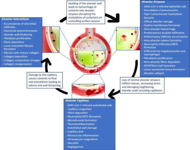
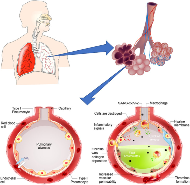
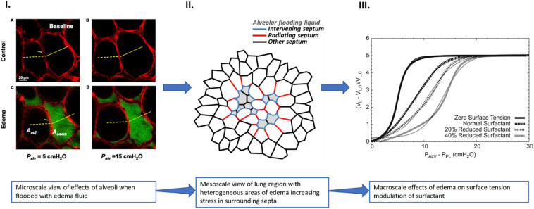
The COVID-19 pandemic surges on as vast research is produced to study the novel SARS-CoV-2 virus and the disease state it induces. Still, little is known about the impact of COVID-19-induced microscale damage in the lung on global lung dynamics. This review summarizes the key histological features of SARS-CoV-2 infected alveoli and links the findings to structural tissue changes and surfactant dysfunction affecting tissue mechanical behavior similar to changes seen in other lung injury. Along with typical findings of diffuse alveolar damage affecting the interstitium of the alveolar walls and blood-gas barrier in the alveolar airspace, COVID-19 can cause extensive microangiopathy in alveolar capillaries that further contribute to mechanical changes in the tissues and may differentiate it from previously studied infectious lung injury. Understanding microlevel damage impact on tissue mechanics allows for better understanding of macroscale respiratory dynamics. Knowledge gained from studies into the relationship between microscale and macroscale lung mechanics can allow for optimized treatments to improve patient outcomes in case of COVID-19 and future respiratory-spread pandemics.

Keywords: Coronavirus infection; Diffuse alveolar damage; Fibrotic lesions; Microangiopathy; Micromechanics; Surfactant dysfunction.


Conflict of interest statement

All authors declare no competing interest.

Figures

Unlabelled Image
Graphical abstract
Fig. 1
Fig. 1
Collagen and elastin fiber network in the lung. I. A. Fluorescence microscopy of thin lung tissue sections stained with Sirius red collagen and green elastin staining concentrated at alveolar septal edges (arrows) (bar = 100 μm) (used with permission from [13]). II. Depiction of forces in the axial, septal and peripheral fiber systems, acting as the stress-bearing elements of the acinar airspaces, (a). Healthy rat lungs fixed at low pressure show narrow alveolar ducts with folded and pleated septal walls of alveoli. Fine dashed lines show the border between the duct and alveolar airspaces, while red and green arrows depict the direction of surface tension force (green) and the counteracting pull of axial system fibers at entrance rings (red). The axial network of collagen and elastin fibers is concentrated at the edges of alveolar septa, coil the ductal airspaces, and form the alveolar entrance rings (represented by the connecting thick gray lines), (c). Healthy rat lungs fixed at higher pressure, showing widening of alveolar ducts and stretching of alveolar septa, with the surface tension force counteracted by the tensile force of axial fiber system in the direction of the ductal lumen, (b, d). Schematic representation of the axial fiber network of collagen and elastin coiling the alveolar duct (depicted by red springs) and stabilizing the alveolar walls with changes in pressure. The axial fiber network is connected to the peripheral fiber system of the pleura (thick black line) through the alveolar septal fibers (green lines), which are found between the basal laminae of the alveolar epithelium and endothelium. Without these three fiber systems, the surface tension would cause the alveoli to collapse. During inflation, pressure gradient due to differences in the pleural pressure (PPl) and alveolar pressure (Palv) induce outward forces (FO) in the peripheral fiber system, which are transmitted into the fiber system of the septal wall (Fi) and eventually are counteracted by the tension in the axial fiber system of the ductal airspace (used from [11], Creative Commons Attribution 4.0 International License, http://creativecommons.org/licenses/by/4.0/). (For interpretation of the references to colour in this figure legend, the reader is referred to the web version of this article.)
Fig. 2
Fig. 2
Flow diagram of study selection.
Fig. 3
Fig. 3
SARS-CoV-2 infection induces alveolar level changes in the lung. The pulmonary alveolus is lined with type I and type II epithelial cells and surrounded by a meshwork of capillaries to facilitate gas exchange. As the healthy alveolus (left) becomes infected with the SARS-CoV-2 virus (right) damage occurs to the alveolar tissues and capillaries. The alveolar damage leads to build-up of debris and fluid inside the alveolus, affecting gas exchange function (images adapted and sourced from shutterstock.com).
Fig. 4
Fig. 4
Microscale damage at the alveolar level leads to formation of stress concentrators affecting lung function at the macroscale. I.) optical sections of alveoli in isolated rat lung show edema formation in individual alveolus causing inflation differences between flooded and aerated alveoli (Aedem = flooded alveolus area, Aadj = aerated alveolus area) A. aerated alveoli at 5 cmH2O alveolar pressure B. inflation of aerated alveoli when alveolar pressure increased to 15 cmH2O C. fluid accumulation in single alveolus causes decrease in size with increase in size of neighboring aerated alveolus at 5 cmH2O alveolar pressure D. when alveolar pressure is increased to 15 cmH2O there is greater difference in inflation between aerated and flooded alveoli (Reprinted with permission of the American Thoracic Society. Copyright © 2020 American Thoracic Society. All rights reserved. Perlman et al. [131]. The American Journal of Respiratory Cell and Molecular Biology is an official journal of the American Thoracic Society [131]). II.) Areas of flooded alveoli may act as stress concentrators increasing stress in septa of surrounding alveoli. When alveoli become flooded and decrease in size, the forces on the walls of neighboring alveoli (radiating septum) increases leading to concentrated areas of increased stress in the lung (adapted from open access [6]). III.) As edema formation impacts the ability of surfactant to control surface tension in multiple focal areas of the lung, global lung function can become affected. The reduction in surface tension control at the microscale can cause a shift in the pressure-volume curve, as shown in silico modeling studies. The graph shows the sigmoid-shaped pressure-volume relationship for a block of 1241 alveoli computed for four different surfactant levels where PALV, PPL, VL and VL0 are the alveolar pressure, pleural pressure, total alveolar volume and total alveolar volume when all collagen and elastin fibers are at rest respectively. The computer model shows that as the amount of surfactant in the lung increases, the volume of alveoli at a fixed pressure increases as well (used with permission from [125]).
Fig. 5
Fig. 5
When the alveoli of the lung become infected with COVID-19, damage occurs that disrupts the alveolar airspace, interstitium and capillaries. Pathological findings that occur within each compartment of the alveoli are summarized. The pathological changes in each compartment disrupt the mechanical behavior of the alveoli which can induce further damage to neighboring areas. In this way, the impacts of COVID-19 on the airspace, interstitium, and capillaries are interconnected and lead to progression of one another (image adapted and sourced from shutterstock.com).

Similar articles

Cited by

References

    1. Dong E., Du H., Gardner L. An interactive web-based dashboard to track COVID-19 in real time. Lancet Infect. Dis. 2020;20:533–534. doi: 10.1016/S1473-3099(20)30120-1. - DOI - PMC - PubMed
    1. Gattinoni L., Chiumello D., Caironi P., Busana M., Romitti F., Brazzi L., Camporota L. COVID-19 pneumonia: different respiratory treatments for different phenotypes? Intensive Care Med. 2020;46:1099–1102. doi: 10.1007/s00134-020-06033-2. - DOI - PMC - PubMed
    1. F.M. Beloncle, B. Pavlovsky, C. Desprez, N. Fage, P.Y. Olivier, P. Asfar, J.C. Richard, A. Mercat, Recruitability and effect of PEEP in SARS-Cov-2-associated acute respiratory distress syndrome, Ann. Intensive Care. 10 (2020). doi:10.1186/s13613-020-00675-7. - DOI - PMC - PubMed
    1. Polak S.B., Van Gool I.C., Cohen D., von der Thüsen J.H., van Paassen J. A systematic review of pathological findings in COVID-19: a pathophysiological timeline and possible mechanisms of disease progression. Mod. Pathol. 2020 doi: 10.1038/s41379-020-0603-3. - DOI - PMC - PubMed
    1. Suki B., Bates J.H.T. Lung tissue mechanics as an emergent phenomenon. J. Appl. Physiol. 2011;110:1111–1118. doi: 10.1152/japplphysiol.01244.2010. - DOI - PMC - PubMed