

Gauging Working Memory Capacity From Differential Resting Brain Oscillations in Older Individuals With A Wearable Device
- PMID: 33716711
- PMCID: PMC7944100
- DOI: 10.3389/fnagi.2021.625006
Gauging Working Memory Capacity From Differential Resting Brain Oscillations in Older Individuals With A Wearable Device
Abstract
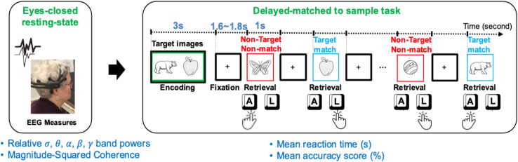
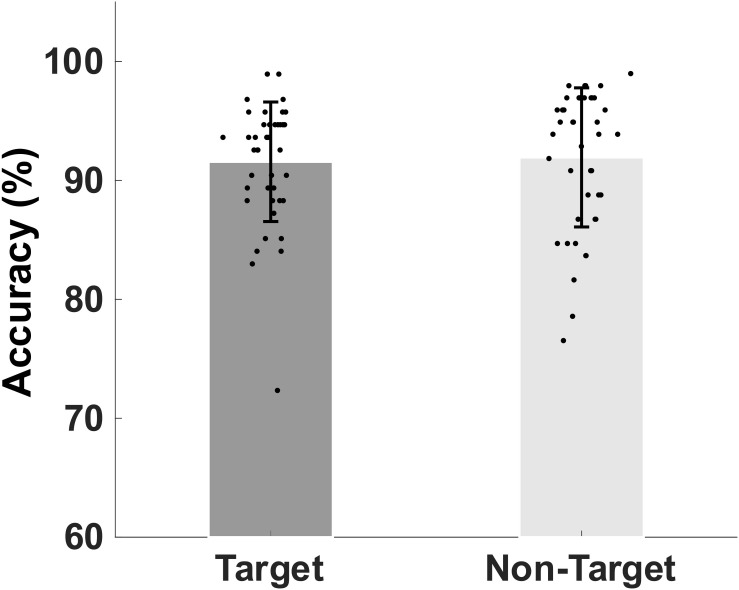
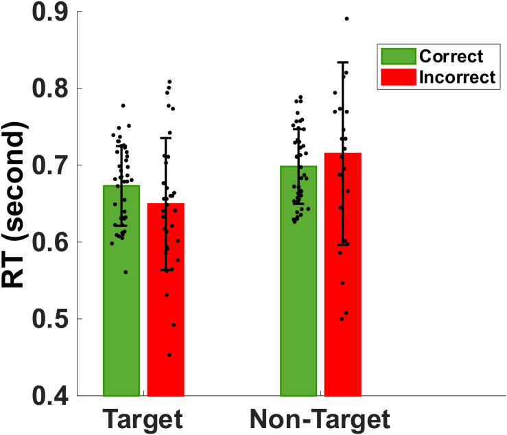
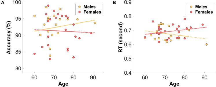
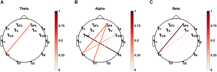
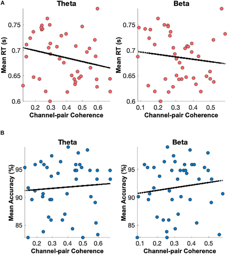
Working memory is a core cognitive function and its deficits is one of the most common cognitive impairments. Reduced working memory capacity manifests as reduced accuracy in memory recall and prolonged speed of memory retrieval in older adults. Currently, the relationship between healthy older individuals' age-related changes in resting brain oscillations and their working memory capacity is not clear. Eyes-closed resting electroencephalogram (rEEG) is gaining momentum as a potential neuromarker of mild cognitive impairments. Wearable and wireless EEG headset measuring key electrophysiological brain signals during rest and a working memory task was utilized. This research's central hypothesis is that rEEG (e.g., eyes closed for 90 s) frequency and network features are surrogate markers for working memory capacity in healthy older adults. Forty-three older adults' memory performance (accuracy and reaction times), brain oscillations during rest, and inter-channel magnitude-squared coherence during rest were analyzed. We report that individuals with a lower memory retrieval accuracy showed significantly increased alpha and beta oscillations over the right parietal site. Yet, faster working memory retrieval was significantly correlated with increased delta and theta band powers over the left parietal sites. In addition, significantly increased coherence between the left parietal site and the right frontal area is correlated with the faster speed in memory retrieval. The frontal and parietal dynamics of resting EEG is associated with the "accuracy and speed trade-off" during working memory in healthy older adults. Our results suggest that rEEG brain oscillations at local and distant neural circuits are surrogates of working memory retrieval's accuracy and processing speed. Our current findings further indicate that rEEG frequency and coherence features recorded by wearable headsets and a brief resting and task protocol are potential biomarkers for working memory capacity. Additionally, wearable headsets are useful for fast screening of cognitive impairment risk.
Keywords: EEG; coherence analysis; mild cognitive impairment; resting-state; working memory.
Copyright © 2021 Borhani, Zhao, Kelly, Gottschalk, Yuan, Jicha and Jiang.
Conflict of interest statement
The authors declare that the research was conducted in the absence of any commercial or financial relationships that could be construed as a potential conflict of interest.
Figures
Similar articles
-
Age-Related Differences in Resting-State EEG and Allocentric Spatial Working Memory Performance.Front Aging Neurosci. 2021 Nov 4;13:704362. doi: 10.3389/fnagi.2021.704362. eCollection 2021. Front Aging Neurosci. 2021. PMID: 34803651 Free PMC article.
-
Study on EEG power and coherence in patients with mild cognitive impairment during working memory task.J Zhejiang Univ Sci B. 2005 Dec;6(12):1213-9. doi: 10.1631/jzus.2005.B1213. J Zhejiang Univ Sci B. 2005. PMID: 16358382 Free PMC article. Clinical Trial.
-
Functional localization and effective connectivity of cortical theta and alpha oscillatory activity during an attention task.Clin Neurophysiol Pract. 2017 Oct 14;2:193-200. doi: 10.1016/j.cnp.2017.09.002. eCollection 2017. Clin Neurophysiol Pract. 2017. PMID: 30214995 Free PMC article.
-
Frontal midline theta oscillations during working memory maintenance and episodic encoding and retrieval.Neuroimage. 2014 Jan 15;85 Pt 2(0 2):721-9. doi: 10.1016/j.neuroimage.2013.08.003. Epub 2013 Aug 8. Neuroimage. 2014. PMID: 23933041 Free PMC article. Review.
-
Music Training, Working Memory, and Neural Oscillations: A Review.Front Psychol. 2020 Feb 21;11:266. doi: 10.3389/fpsyg.2020.00266. eCollection 2020. Front Psychol. 2020. PMID: 32153474 Free PMC article. Review.
Cited by
-
Parallel electrophysiological abnormalities due to COVID-19 infection and to Alzheimer's disease and related dementia.Alzheimers Dement. 2024 Oct;20(10):7296-7319. doi: 10.1002/alz.14089. Epub 2024 Aug 29. Alzheimers Dement. 2024. PMID: 39206795 Free PMC article.
-
Physical exercise for brain plasticity promotion an overview of the underlying oscillatory mechanism.Front Neurosci. 2024 Aug 8;18:1440975. doi: 10.3389/fnins.2024.1440975. eCollection 2024. Front Neurosci. 2024. PMID: 39176382 Free PMC article. Review.
-
Distinct Effects of the Apolipoprotein E ε4 Genotype on Associations Between Delayed Recall Performance and Resting-State Electroencephalography Theta Power in Elderly People Without Dementia.Front Aging Neurosci. 2022 May 26;14:830149. doi: 10.3389/fnagi.2022.830149. eCollection 2022. Front Aging Neurosci. 2022. PMID: 35693343 Free PMC article.
-
Screening for Mild Cognitive Impairment with Speech Interaction Based on Virtual Reality and Wearable Devices.Brain Sci. 2023 Aug 21;13(8):1222. doi: 10.3390/brainsci13081222. Brain Sci. 2023. PMID: 37626578 Free PMC article.
-
Dissociable neural mechanisms of cognition and well-being in youth versus healthy aging.Psychol Aging. 2022 Nov;37(7):827-842. doi: 10.1037/pag0000710. Epub 2022 Sep 15. Psychol Aging. 2022. PMID: 36107693 Free PMC article.
References
-
- Abiri R., Borhani S., Jiang Y., Zhao X. (2019a). Decoding attentional state to faces and scenes using EEG brainwaves. Complexity 2019:6862031.
-
- App-LabRecorder (2019). Available online at: https://github.com/labstreaminglayer/App-LabRecorder (accessed 8/10/2019).
Grants and funding
LinkOut - more resources
Full Text Sources
Other Literature Sources