

Cerebrovascular Reactivity Measurement Using Magnetic Resonance Imaging: A Systematic Review
- PMID: 33716793
- PMCID: PMC7947694
- DOI: 10.3389/fphys.2021.643468
Cerebrovascular Reactivity Measurement Using Magnetic Resonance Imaging: A Systematic Review
Erratum in
-
Corrigendum: Cerebrovascular reactivity measurement using magnetic resonance imaging: A systematic review.Front Physiol. 2022 Dec 9;13:1105285. doi: 10.3389/fphys.2022.1105285. eCollection 2022. Front Physiol. 2022. PMID: 36569753 Free PMC article.
Abstract
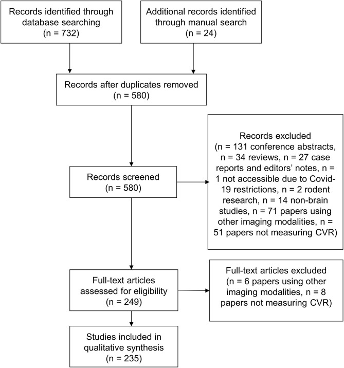
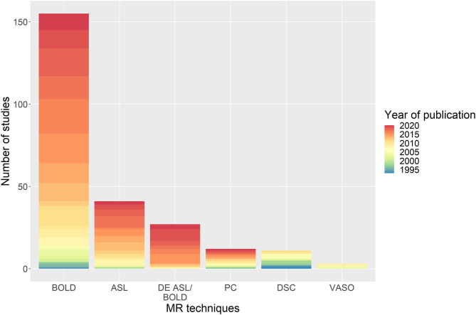
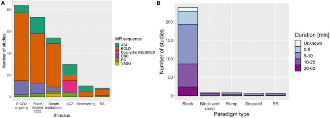
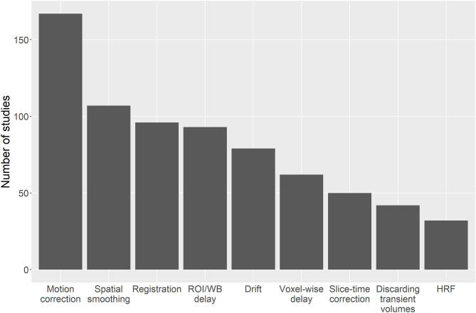
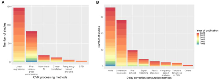
Cerebrovascular reactivity (CVR) magnetic resonance imaging (MRI) probes cerebral haemodynamic changes in response to a vasodilatory stimulus. CVR closely relates to the health of the vasculature and is therefore a key parameter for studying cerebrovascular diseases such as stroke, small vessel disease and dementias. MRI allows in vivo measurement of CVR but several different methods have been presented in the literature, differing in pulse sequence, hardware requirements, stimulus and image processing technique. We systematically reviewed publications measuring CVR using MRI up to June 2020, identifying 235 relevant papers. We summarised the acquisition methods, experimental parameters, hardware and CVR quantification approaches used, clinical populations investigated, and corresponding summary CVR measures. CVR was investigated in many pathologies such as steno-occlusive diseases, dementia and small vessel disease and is generally lower in patients than in healthy controls. Blood oxygen level dependent (BOLD) acquisitions with fixed inspired CO2 gas or end-tidal CO2 forcing stimulus are the most commonly used methods. General linear modelling of the MRI signal with end-tidal CO2 as the regressor is the most frequently used method to compute CVR. Our survey of CVR measurement approaches and applications will help researchers to identify good practice and provide objective information to inform the development of future consensus recommendations.
Keywords: Hypercapnia (CO(2)) inhalation; arterial spin labelling MRI; blood oxygen-level dependent; cerebrovascular reactivity; magnetic resonance imaging; systematic review.
Copyright © 2021 Sleight, Stringer, Marshall, Wardlaw and Thrippleton.
Conflict of interest statement
The authors declare that the research was conducted in the absence of any commercial or financial relationships that could be construed as a potential conflict of interest.
Figures
References
-
- Atwi S., Shao H., Crane D. E., da Costa L., Aviv R. I., Mikulis D. J., et al. . (2019). BOLD-based cerebrovascular reactivity vascular transfer function isolates amplitude and timing responses to better characterize cerebral small vessel disease. NMR Biomed. 32:e4064. 10.1002/nbm.4064 - DOI - PubMed
Publication types
Grants and funding
LinkOut - more resources
Full Text Sources
Other Literature Sources