. 2021 Feb 25:12:647289.
doi: 10.3389/fmicb.2021.647289. eCollection 2021.

New Insights Into the Antibacterial Mechanism of Cryptotanshinone, a Representative Diterpenoid Quinone From Salvia miltiorrhiza Bunge

Affiliations

New Insights Into the Antibacterial Mechanism of Cryptotanshinone, a Representative Diterpenoid Quinone From Salvia miltiorrhiza Bunge

Bo-Chen Chen et al. Front Microbiol. .

Abstract

The rapid rise of antibiotic resistance causes an urgent need for new antimicrobial agents with unique and different mechanisms of action. The respiratory chain is one such target involved in the redox balance and energy metabolism. As a natural quinone compound isolated from the root of Salvia miltiorrhiza Bunge, cryptotanshinone (CT) has been previously demonstrated against a wide range of Gram-positive bacteria including multidrug-resistant pathogens. Although superoxide radicals induced by CT are proposed to play an important role in the antibacterial effect of this agent, its mechanism of action is still unclear. In this study, we have shown that CT is a bacteriostatic agent rather than a bactericidal agent. Metabolome analysis suggested that CT might act as an antibacterial agent targeting the cell membrane. CT did not cause severe damage to the bacterial membrane but rapidly dissipated membrane potential, implying that this compound could be a respiratory chain inhibitor. Oxygen consumption analysis in staphylococcal membrane vesicles implied that CT acted as respiratory chain inhibitor probably by targeting type II NADH:quinone dehydrogenase (NDH-2). Molecular docking study suggested that the compound would competitively inhibit the binding of quinone to NDH-2. Consistent with the hypothesis, the antimicrobial activity of CT was blocked by menaquinone, and the combination of CT with thioridazine but not 2-n-heptyl-4-hydroxyquinoline-N-oxide exerted synergistic activity against Staphylococcus aureus. Additionally, combinations of CT with other inhibitors targeting different components of the bacterial respiratory chain exhibit potent synergistic activities against S. aureus, suggesting a promising role in combination therapies.

Keywords: cryptotanshinone; menaquinone; metabolome analysis; respiratory chain inhibitor; type II NADH:quinone dehydrogenase.


Conflict of interest statement

The authors declare that the research was conducted in the absence of any commercial or financial relationships that could be construed as a potential conflict of interest.

Figures
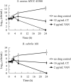
FIGURE 1
FIGURE 1
Time-kill curves for Bacillus subtilis and Staphylococcus aureus treated with CT. The curves are viable cell concentrations plotted against time. MIC, minimum inhibitory concentration; VAN, vancomycin. DMSO (3.2%) is as a no drug control. Data represent the mean ± SD (n = 3).
FIGURE 2
FIGURE 2
CT significantly affects the glycerophospholipid metabolism of Bacillus subtilis. DMSO is as a control without drug (Con). Deep red represents the highest level, and deep blue represents the lowest level, with white representing equal levels. Each group is identified by green (CT) and red (DMSO) bars at the top of each column. Six biological replicates were tested for each sample.
FIGURE 3
FIGURE 3
Effect of CT on bacterial membrane integrity. (A) Determination of cellular element concentrations of B. subtilis 168 by inductively coupled plasma optical emission spectroscopy after CT treatment for 30 min. DMSO, non-drug treated control; VAN (vancomycin), negative control; Nig (nigericin), specific potassium ionophore; CCCP, depolarization agent. The experiments were carried out in triplicate in two independent replications. (B) Leakage of ATP following exposure of B. subtilis 168 to antimicrobial agents. Nisin, a pore-forming agent. Data are presented as mean ± SD of three independent experiments with triplicate measurements. (C) Staining with propidium iodide. Representative data from three independent cultures of S. aureus ATCC 43300 and B. subtilis 168 were shown following exposure to antimicrobial agents at 8 × MIC for 120 min.
FIGURE 4
FIGURE 4
Effect of CT on membrane potential of B. subtilis 168 (A,C) and S. aureus ATCC 43300 (B,D). (A,B) Membrane potential estimation by flow cytometry using DiOC2(3) dye; Representative data from three independent cultures of both strains were shown following exposure to different agents for 10 min. (C,D) Fluorescence of DiOC2(3) was detected at an excitation wavelength of 485 nm and two emission wavelengths, 530 nm (green) and 630 nm (red), using a microplate reader. Addition of CT to the cell suspensions of bacteria is indicated by an arrow. Data are presented as mean ± SD of three independent experiments. DMSO (1.6%), non-drug treated control; CCCP, depolarization control; Nigericin, hyperpolarization control; VAN (vancomycin), negative control.
FIGURE 5
FIGURE 5
The effects of CT on respiratory chain activity of bacteria. (A–D) Inhibition of oxygen consumption of staphylococcal membrane vesicles by CT. Respiration was initiated by the addition of 1 mM NADH. Thioridazine (THZ) was used as positive control. Vancomycin (VAN) and DMSO were used as negative control, respectively. CT, 80 μg/mL; THZ, 100 μg/mL; VAN, 64 μg/mL; DMSO, 2%; dithiothreitol (DTT), 2.85 mM; ubiquinone-10 (Q10), 40 μM. (E) Effect of CT on NADH/NAD+ ratios. Intracellular NADH/NAD+ ratios of S. aureus ATCC 43300 were determined by an Amplite fluorimetric NAD/NADH ratio assay kit. DMSO, 3.2%; CT, 32 μg/mL; thioridazine (THZ), 64 μg/mL; HQNO, 32 μg/mL.
FIGURE 6
FIGURE 6
CT competitively inhibits the binding of quinone to NDH-2. (A) Configuration for the interaction of ubiquinone-5 (UQ5) with NDH-2. (B) Configuration for the interaction of CT with NDH-2. (C) MK4 antagonizes the antibacterial activity of CT.
FIGURE 7
FIGURE 7
Effect of CT on ATP synthesis. (A) Cellular ATP levels in B. subtilis 168 were measured in the presence of CT at specified concentration after the addition of 10 mM glucose for 30 min. Vancomycin (VAN, 2 μg/mL) and CCCP (8 μg/mL) were used as negative and positive control, respectively. (B) The ATP synthesis activity was determined in membrane vesicles isolated from S. aureus ATCC 43300 energized with NADH. Vancomycin (VAN, 32 μg/mL) and Tomatidine (TO, 64 μg/mL) were used as negative and positive control, respectively. Data from three independent experiments are presented as mean ± SD.

Similar articles

Cited by

References

    1. Allen N. E., Alborn W. E., Hobbs J. N. (1991). Inhibition of membrane potential-dependent amino acid transport by daptomycin. Antimicrob. Agents Chemother. 35 2639–2642. 10.1128/AAC.35.12.2639 - DOI - PMC - PubMed
    1. Al-Mebairik N. F., El-Kersh T. A., Al-Sheikh Y. A., Marie M. A. M. (2016). A review of virulence factors, pathogenesis, and antibiotic resistance in Staphylococcus aureus. Rev. Med. Microbiol. 27 50–56. 10.1097/MRM.0000000000000067 - DOI
    1. Andries K. (2005). A Diarylquinoline Drug Active on the ATP Synthase of Mycobacterium tuberculosis. Science 307 223–227. 10.1126/science.1106753 - DOI - PubMed
    1. Anraku Y. (1988). Bacterial Electron Transport Chains. Annu. Rev. Biochem. 57 101–132. 10.1146/annurev.bi.57.070188.000533 - DOI - PubMed
    1. Azarkina N., Siletsky S., Borisov V., von Wachenfeldt C., Hederstedt L., Konstantinov A. A. (1999). A Cytochrome bb ‘-type Quinol Oxidase in Bacillus subtilis Strain 168. J. Biol. Chem. 274 32810–32817. 10.1074/jbc.274.46.32810 - DOI - PubMed

LinkOut - more resources