

Triple-Negative Breast Cancer: Intact Mismatch Repair and Partial Co-Expression of PD-L1 and LAG-3
- PMID: 33717059
- PMCID: PMC7943629
- DOI: 10.3389/fimmu.2021.561793
Triple-Negative Breast Cancer: Intact Mismatch Repair and Partial Co-Expression of PD-L1 and LAG-3
Abstract
Background and aim: Poor response to immune checkpoint inhibitors (ICIs) has been observed in most triple-negative breast cancer (TNBC) cases (around 80%). Our aim was to investigate the status of mismatch repair (MMR), microsatellite instability (MSI), programmed death-ligand 1 (PD-L1), and lymphocyte-activation gene 3 (LAG-3) in TNBC.
Methods: A total of 74 TNBC samples were retrospectively analyzed. MMR and MSI were evaluated by immunohistochemistry (IHC) and polymerase chain reaction (PCR) using Promega 1.2 and NCI panels, respectively. PD-L1, LAG-3, and CD8 expression was assessed by IHC.
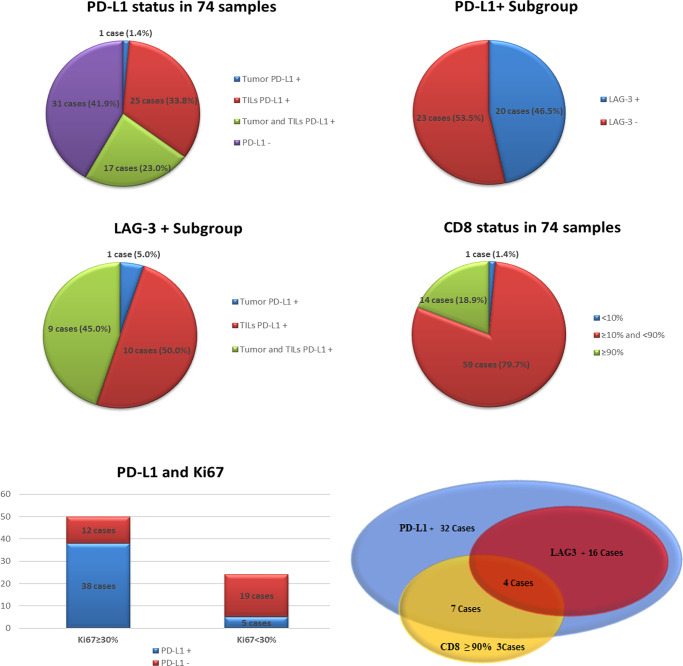
Results: None of the cases demonstrated deficient MMR (dMMR) or MSI. In total, 43/74 cases (58.1%) were PD-L1+, including 1 tumor PD-L1+, 25 tumor-infiltrating lymphocytes (TILs) PD-L1+, and 17 cases involving concurrence of tumor and TIL PD-L1+. The rate of TIL PD-L1+ was remarkably higher than that of tumor PD-L1+ (P<0.001). We identified 20 LAG-3+ cases (27.0%, 20/74), all of which were PD-L1+. Co-expression of PD-L1 and LAG-3 was noted in 46.5% (20/43) of the PD-L1+ population. In the LAG-3+ subtype (co-expression of PD-L1 and LAG-3), high correlation between TILs PD-L1+ and LAG-3+ was observed (P<0.01). A high frequency of CD8+ (98.6%, 73/74) was observed.
Conclusion: dMMR/MSI characteristics may not be a practical predictive marker for ICIs in TNBC. PD-L1+ is more common in TILs than in tumors. In the PD-L1+ population, approximately half of the cases showed LAG-3 co-expression. For patients with a poor response to PD-1(L1) mono ICI, dual blockade of PD-1(L1) and LAG-3 may be a viable option for the management of TNBC.
Keywords: CD8; LAG-3; PD-L1; microsatellite instability; triple-negative breast cancer.
Copyright © 2021 Wu, Shi, Wang, Wang, Liu, Luo, Mao and Zeng.
Conflict of interest statement
The authors declare that the research was conducted in the absence of any commercial or financial relationships that could be construed as a potential conflict of interest.
Figures


Similar articles
-
Microsatellite instability and mismatch repair protein expressions in lymphocyte-predominant breast cancer.Cancer Sci. 2020 Jul;111(7):2647-2654. doi: 10.1111/cas.14500. Epub 2020 Jun 13. Cancer Sci. 2020. PMID: 32449246 Free PMC article.
-
Can evaluation of mismatch repair defect and TILs increase the number of triple-negative breast cancer patients eligible for immunotherapy?Pathol Res Pract. 2021 Oct;226:153606. doi: 10.1016/j.prp.2021.153606. Epub 2021 Aug 31. Pathol Res Pract. 2021. PMID: 34530255
-
Mismatch repair protein deficiency in triple-negative breast carcinomas.J Int Med Res. 2024 Jun;52(6):3000605241259747. doi: 10.1177/03000605241259747. J Int Med Res. 2024. PMID: 38902203 Free PMC article.
-
ESMO recommendations on microsatellite instability testing for immunotherapy in cancer, and its relationship with PD-1/PD-L1 expression and tumour mutational burden: a systematic review-based approach.Ann Oncol. 2019 Aug 1;30(8):1232-1243. doi: 10.1093/annonc/mdz116. Ann Oncol. 2019. PMID: 31056702
-
Microsatellite Instability, Mismatch Repair, and Tumor Mutation Burden in Lung Cancer.Surg Pathol Clin. 2024 Jun;17(2):295-305. doi: 10.1016/j.path.2023.11.011. Epub 2023 Dec 20. Surg Pathol Clin. 2024. PMID: 38692812 Review.
Cited by
-
LAG-3 as a Potent Target for Novel Anticancer Therapies of a Wide Range of Tumors.Int J Mol Sci. 2022 Sep 1;23(17):9958. doi: 10.3390/ijms23179958. Int J Mol Sci. 2022. PMID: 36077354 Free PMC article. Review.
-
Prevalence of Epstein-Barr Virus Infection and Mismatch Repair Protein Deficiency and the Correlation of Immune Markers in Tibetan Patients with Gastric Cancer.Biomed Res Int. 2022 Jun 13;2022:2684065. doi: 10.1155/2022/2684065. eCollection 2022. Biomed Res Int. 2022. PMID: 35734348 Free PMC article.
-
Immune Checkpoint LAG3 and Its Ligand FGL1 in Cancer.Front Immunol. 2022 Jan 17;12:785091. doi: 10.3389/fimmu.2021.785091. eCollection 2021. Front Immunol. 2022. PMID: 35111155 Free PMC article. Review.
-
Herpesvirus entry mediator as a potential biomarker in breast cancer compared with conventional cytotoxic T‑lymphocyte‑associated antigen 4.Biomed Rep. 2023 Jul 17;19(2):56. doi: 10.3892/br.2023.1638. eCollection 2023 Aug. Biomed Rep. 2023. PMID: 37560313 Free PMC article.
-
DNA Mismatch Repair System Imbalances in Breast Adenocarcinoma.Cancer Diagn Progn. 2023 Mar 3;3(2):169-174. doi: 10.21873/cdp.10197. eCollection 2023 Mar-Apr. Cancer Diagn Progn. 2023. PMID: 36875308 Free PMC article. Review.
References
Publication types
MeSH terms
Substances
LinkOut - more resources
Full Text Sources
Other Literature Sources
Research Materials