Review
. 2021 Feb 25:12:600829.
doi: 10.3389/fimmu.2021.600829. eCollection 2021.

Biomechanics of T Cell Dysfunctions in Chronic Diseases

Affiliations
Review

Biomechanics of T Cell Dysfunctions in Chronic Diseases

Sachith D Gunasinghe et al. Front Immunol. .

Abstract

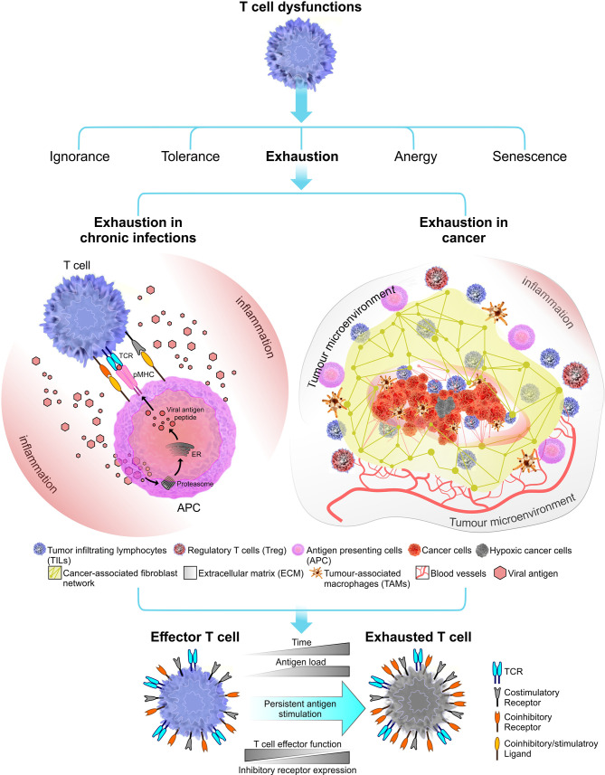
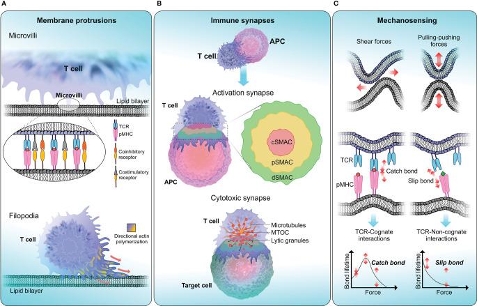
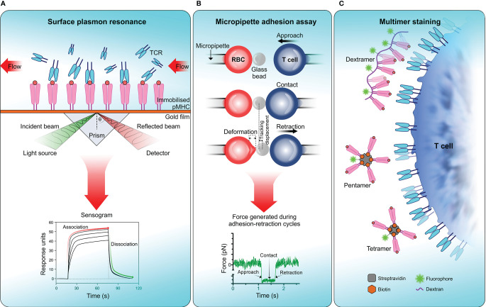
Understanding the mechanisms behind T cell dysfunctions during chronic diseases is critical in developing effective immunotherapies. As demonstrated by several animal models and human studies, T cell dysfunctions are induced during chronic diseases, spanning from infections to cancer. Although factors governing the onset and the extent of the functional impairment of T cells can differ during infections and cancer, most dysfunctional phenotypes share common phenotypic traits in their immune receptor and biophysical landscape. Through the latest developments in biophysical techniques applied to explore cell membrane and receptor-ligand dynamics, we are able to dissect and gain further insights into the driving mechanisms behind T cell dysfunctions. These insights may prove useful in developing immunotherapies aimed at reinvigorating our immune system to fight off infections and malignancies more effectively. The recent success with checkpoint inhibitors in treating cancer opens new avenues to develop more effective, targeted immunotherapies. Here, we highlight the studies focused on the transformation of the biophysical landscape during infections and cancer, and how T cell biomechanics shaped the immunopathology associated with chronic diseases.

Keywords: T cell dysfunction; biomechanics; biophysical landscape; cancer; chronic diseases; immune receptor landscape; infections; tumor microenvironment.


Conflict of interest statement

The authors declare that the research was conducted in the absence of any commercial or financial relationships that could be construed as a potential conflict of interest.

Figures

Figure 1
Figure 1
Immune receptor landscape during T cell exhaustion. Exhaustion can be induced by chronic infections (in this instant viral infections) or cancer. Factors that influence the onset and the extent of T cell exhaustion differ in these two exhaustion models. During a chronic infection, pathogen clearance become inefficient, leading to persistent inflammation and chronic antigen stimulation of T cells which results in clonal deletion or exhaustion. In cancer, the immunosuppressive tumor microenvironment plays a crucial role in shaping the outcome of T cell exhaustion. Tumor microenvironment comprised of stroma containing a fibroblast network and a number of immune cells including regulatory T cells (Tregs) and tumor-associated macrophages (TAMs) which together promote tumorigenesis (77). Tumor microenvironment can induce stromal cells to secrete growth factor to promote angiogenesis (i.e. grow new blood vessels that feed the tumor) (78). Overall, T cell exhaustion in both chronic infections and cancer known to have several overlapping functional and phenotypic characteristics. The most common feature is sustained upregulation of inhibitory receptors during the course of the disease.
Figure 2
Figure 2
T cell biophysical landscape . (A) Membrane protrusions. T cells continuously form actin-rich membrane protrusions known as filopodia or microvilli which help them to sense biophysical properties in the surrounding environment. Microvilli are involved in early T cell activation (173) and where signaling components including TCR and TCR-associated signaling molecules get accumulated (174, 175). The tip of a microvilli is zoomed in to illustrate the accumulation of T cell signaling molecules. Compared to microvilli, filopodium membrane projections are larger in size ranging from 10-40 µm in length in different cells (176). The function of filopodia is broad including crucial roles in cell-cell adhesion and cell migration (177). (B) Immune synapses. Formation of immune synapses are important steps in T cell activation and executing T cell effector functions through cytotoxicity. When forming activation synapses, signaling receptors (in T cells) and ligands (in APCs) spatially segregate into a bull’s eye-like structure forming the supra-molecular activation cluster that is separated into central (cSMAC—red), peripheral (pSMAC—yellow) and distal (dSMAC—green) regions, where each zone preferentially recruit different signaling receptors to initiate T cell signaling. Cytotoxic synapses are formed when a CTL encounters a target cell. Cytotoxic synapses differ from activation synapses in the recruitment of lytic granules to the synaptic cleft with the help of Golgi apparatus and the microtubule organising centre (MTOC). Delivery of lytic granules and exocytosis of granule contents are highly depended on calcium influx. (C) Mechanosensing. T cells are constantly being subjected to mechanical stresses when undergoing kinapse formation with APCs. TCRs display mechanosensing properties by exerting pulling-pushing and shear forces on pMHC molecules on APCs. When a TCR engage with cognate-antigen peptide, a catch bond is formed, which triggers conformational changes in the TCR-CD3 complex and initiate T cell signaling. Conversely, TCR encounter with a non-cognate-antigen peptide results in a slip bond, where an exponential decay of bond lifetime is observed with increasing force. Slip bonds fail to trigger TCR signaling.
Figure 3
Figure 3
Measuring TCR affinity . (A) Surface plasmon resonance (SPR). SPR measures the equilibrium dissociation constant (KD) of TCR-pMHC interactions in which pMHC is immobilized on a sensor surface and TCR molecules are injected in a continuous flow. Binding of TCR to pMHC results in a change of mass on the sensor surface and is recorded in a sensogram which is then used to calculate KD. (B) Micropipette adhesion assay. This technique uses two probes, one that is stationary which contains a red blood cell (RBC) attached to a functionalized glass bead to act as the adhesion force transducer and a mobile force probe bearing a T cell coupled to a piezotranslator. During adhesion-retraction cycles carried out by the mobile probe, the deformation of the RBC, displacement of the glass bead and the force generated in each cycle is recorded. (C) Multimer staining. This technique enhances the binding avidity of TCR-pMHC by increasing the valency of the interaction, results in more stable multimeric TCR-pMHC complexes for efficient labelling and detection. To date numerous forms of pMHC multimers have been reported which includes tetramers, pentamers, octamers, and dextramers (289).

References

    1. Rock KL, Reits E, Neefjes J. Present Yourself! By MHC Class I and MHC Class II Molecules. Trends Immunol (2016) 37:724–37. 10.1016/j.it.2016.08.010 - DOI - PMC - PubMed
    1. Natarajan K, Jiang J, May NA, Mage MG, Boyd LF, McShan AC, et al. . The role of molecular flexibility in antigen presentation and T cell receptor-mediated signaling. Front Immunol (2018) 9:1657. 10.3389/fimmu.2018.01657 - DOI - PMC - PubMed
    1. Masopust D, Schenkel JM. The integration of T cell migration, differentiation and function. Nat Rev Immunol (2013) 13:309–20. 10.1038/nri3442 - DOI - PubMed
    1. Cox MA, Harrington LE, Zajac AJ. Cytokines and the inception of CD8 T cell responses. Trends Immunol (2011) 32:180–6. 10.1016/j.it.2011.01.004 - DOI - PMC - PubMed
    1. Kalia V, Sarkar S, Ahmed R. CD8 T-cell memory differentiation during acute and chronic viral infections. Adv Exp Med Biol (2010) 684:79–95. 10.1007/978-1-4419-6451-9_7 - DOI - PubMed

Publication types