Review
. 2021 Feb 26:12:620284.
doi: 10.3389/fimmu.2021.620284. eCollection 2021.

Resident Innate Immune Cells in the Cornea

Affiliations
Review

Resident Innate Immune Cells in the Cornea

Jun Liu et al. Front Immunol. .

Abstract

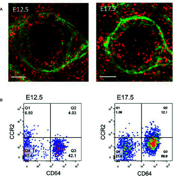
The cornea is a special interface between the internal ocular tissue and the external environment that provides a powerful chemical, physical, and biological barrier against the invasion of harmful substances and pathogenic microbes. This protective effect is determined by the unique anatomical structure and cellular composition of the cornea, especially its locally resident innate immune cells, such as Langerhans cells (LCs), mast cells (MCs), macrophages, γδ T lymphocytes, and innate lymphoid cells. Recent studies have demonstrated the importance of these immune cells in terms of producing different cytokines and other growth factors in corneal homeostasis and its pathologic conditions. This review paper briefly describes the latest information on these resident immune cells by specifically analyzing research from our laboratory.

Keywords: Langerhans cells; cornea; immune cells; innate lymphoid cells; macrophages; mast cells; γδ T-cells.


Conflict of interest statement

The authors declare that the research was conducted in the absence of any commercial or financial relationships that could be construed as a potential conflict of interest.

Figures

Figure 1
Figure 1
Langerhans cells located in the basal layer of the corneal limbal epithelium. (A) Anterior segment of murine eyeball; (B) Langerhans cells (phycoerythrin (PE)-conjugated anti-mouse CD11c staining, red) and basal epithelial cells (4’,6-diamidino-2-phenylindole (DAPI) staining, blue) in the murine corneal limbus. Scale bar: 25 μm.
Figure 2
Figure 2
Dynamic changes in first-wave mast cells (MCs) during the embryonic and neonatal periods. (A) Immunostaining of eyeball cross section with fluorescein isothiocyanate (FITC)-conjugated avidin for mast cells and DAPI for cell nuclei. Scale bars: left and upper-right images, 20 μm; lower-right image, 100 μm. (B) These images depict immunostaining of the cornea with a complete limbus with anti-mouse CD31-PE (red) for blood vessels and FITC-conjugated avidin (green) for mast cells during different developmental stages from E14.5, P13, to Adult. Scale bars: 200 μm. E, embryonic day; P, postnatal day. [From Liu J et al. (67)]. (C) Analysis of the roles of MCs in corneal innervation. The left two images show the immunostaining of the cornea with NL557-conjugated anti-β-III tubulin and Avidin-FITC staining at P1. Scale bars: 200 μ m. The right image shows the differences in nerve fiber density between WT and c-Kit-/- murine corneas. (D) Analysis of the roles of MCs in limbal vasculogenesis. The left two images show the limbal vessel and MC immunostaining of the cornea with anti-mouse CD31-FITC and Avidin-PE staining, respectively. Scale bars: 200 μm. The right image shows the difference in limbal vessel network area between WT and c-Kit-/- murine corneas.
Figure 3
Figure 3
Analysis of corneal macrophages at E12.5 and E17.5. (A) Represented images of macrophage (PE-conjugated anti-mouse CD64+, red) distribution and limbal vessels (fluorescein isothiocyanate (FITC)-conjugated anti-mouse CD31, green) in E12.5 and E17.5 corneas. Scale bars: 200 μm [From Liu J et al. (86)]. (B) The cells in the flow dot plot of E12.5 and E17.5 were both derived from CD45 positive cells. CD64 antigen was used to identify macrophages, and CCR2 antigen was used to distinguished the distinct cell population in corneal macrophages.
Figure 4
Figure 4
Distribution of γδ T-cells in the corneal limbus and conjunctiva. (A) Anterior segment of murine eyeball; (B) γδ T-cells (PE-conjugated anti-mouse TCRγδ, Clone GL3, red) and epithelial cells (DAPI staining, blue) in the murine corneal limbus. Scale bar: 25 μm.
Figure 5
Figure 5
Identification of NK cells and ILC2s in the cornea. (A–F) Immunostaining and phenotypical identification of corneal limbal NK cells with anti-mouse NKp46-FITC, CD3-PE, EOMES-PE, IL-22-PE, RORγt-APC, and CD94-PE in the injured cornea at 24 h after corneal abrasion. (From Liu Q et al. (125). (G) The lineage antibody is a cocktail of anti-mouse CD3, anti-mouse Ly-6G/Ly-6C, anti-mouse CD11b, anti-mouse CD45R/B220, and anti-mouse TER-119 antibodies. ILC2s were identified as a CD45+Lin-CD90.2+CD127+T1/ST2+ cell population. (H) After corneal epithelial wounding, anti-CD90.2 antibody–treated mice were injected i.v. with sorted lung ILC2s stained by carboxyfluorescein diacetate succinimidyl ester. Corneas of those mice stained with DAPI were visualized using fluorescence microscopy 24 h after injection. The red lines represent the limbal vessel wall. Scale bars: 100 μm. (From Liu J et al. (126).

References

    1. Veiga-Fernandes H, Mucida D. Neuro-Immune Interactions at Barrier Surfaces. Cell (2016) 165:801–11. 10.1016/j.cell.2016.04.041 - DOI - PMC - PubMed
    1. Chesne J, Cardoso V, Veiga-Fernandes H. Neuro-immune regulation of mucosal physiology. Mucosal Immunol (2019) 12:10–20. 10.1038/s41385-018-0063-y - DOI - PubMed
    1. Kabashima K, Honda T, Ginhoux F, Egawa G. The immunological anatomy of the skin. Nat Rev Immunol (2019) 19:19–30. 10.1038/s41577-018-0084-5 - DOI - PubMed
    1. Nielsen MM, Witherden DA, Havran WL. γδ T cells in homeostasis and host defence of epithelial barrier tissues. Nat Rev Immunol (2017) 17:733–45. 10.1038/nri.2017.101 - DOI - PMC - PubMed
    1. Powell N, Walker MM, Talley NJ. The mucosal immune system: master regulator of bidirectional gut-brain communications. Nat Rev Gastroenterol Hepatol (2017) 14:143–59. 10.1038/nrgastro.2016.191 - DOI - PubMed

Publication types

LinkOut - more resources