Clinical Trial
. 2021 Feb 26:12:630096.
doi: 10.3389/fimmu.2021.630096. eCollection 2021.

Club Cell Loss as a Feature of Bronchiolization in ILD

Affiliations
Clinical Trial

Club Cell Loss as a Feature of Bronchiolization in ILD

Paul Reynaud et al. Front Immunol. .

Abstract

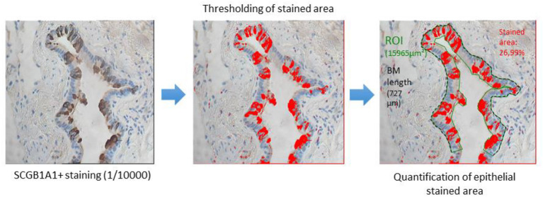
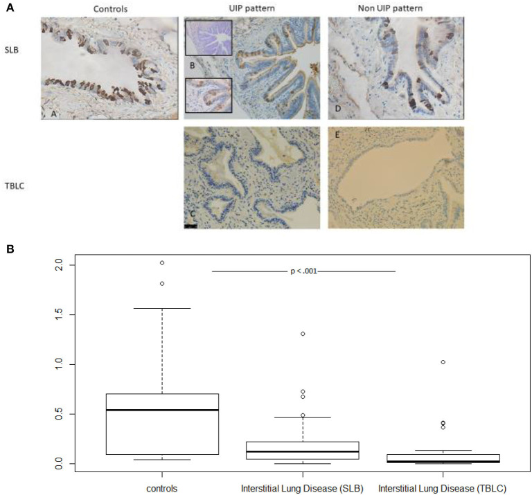
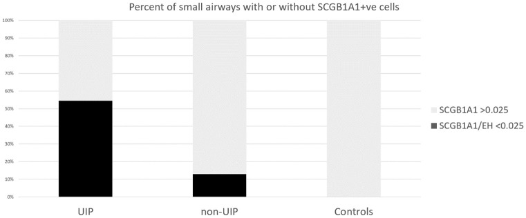
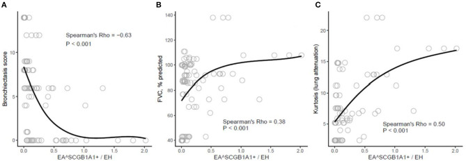
Background: Distal airway metaplasia may precede honeycombing in progressive fibrosing interstitial lung disease (ILD). The SCGB1A1+ bronchiolar-specific club cell may play a role in this aberrant regenerative process. Objective: To assess the presence of club cells in the small airways of patients suffering from ILD. Methods: Small airways (internal diameter <2 mm) in lung samples [surgical lung biopsy (SLB) and/or transbronchial lung cryobiopsy (TBLC)] from 14 patients suffering from ILD and 10 controls were morphologically assessed and stained for SCGB1A1. SCGB1A1 was weighted by epithelial height as a marker of airway generation (SCGB1A1/EH). Correlations between clinical, functional, and high-resolution CT (HRCT) prognostic factors and histomorphometry were assessed. Results: Small airways from samples with ILD patterns were significantly less dense in terms of SCGB1A1+ cells [0.064 (0.020-0.172)] as compared to controls' sample's small airways [0.393 (0.082-0.698), p < 0.0001]. Usual interstitial pneumonia (UIP) patterns most frequently contained small airways with limited or absent SCGB1A1 expression (SCGB1A1/EH <0.025): UIP (18/33; 55%) as compared with non-UIP patterns (4/31; 13%) or controls (0/29; 0%): p < 0.0001. In addition, correlations with HRCT indicated a significant negative relationship between SCGB1A1 and bronchiectasis as a feature of bronchiolization (Rho -0.63, p < 0.001) and a positive relationship with both forced vital capacity (FVC) and Hounsfield unit (HU)-distribution pattern in kurtosis (Rho 0.38 and 0.50, respectively, both p < 0.001) as markers of fibrotic changes. Conclusion: Compared with controls, the small airways of patients with ILD more often lack SCGB1A1, especially so in UIP. Low densities of SCGB1A1-marked cells correlate with bronchiectasis and fibrotic changes. Further research investigating SCGB1A1 staining as a pathological feature of the bronchiolization process is merited.

Keywords: SCGB1A1; bronchiolization; club (clara) cell; idiopathic pulmonary fibrosis; interstitial lung disease; metaplasia.


Conflict of interest statement

The authors declare that the research was conducted in the absence of any commercial or financial relationships that could be construed as a potential conflict of interest.

Figures

Figure 1
Figure 1
After SCGB1A1 staining, small airway epithelia were isolated via thresholding, perimeters, thickness/height, and by the area quantified.
Figure 2
Figure 2
(A) Small airways are immune-stained for SCGB1A1 in controls, UIP, and non-UIP patterns. Upper left: (A) SLB from a control (no TBLC were available in this group). Middle row: (B) Very limited staining in some (left lower part) but not all small airways sampled by SLB; Inserts are: Serial cut with a Blue Alcian staining suggestive of the presence of mucin-costaining and a higher magnification suggestive of goblet cell morphology for SCGB1A1 positively stained epithelial cells. Ectasic small airway from a TBLC UIP pattern with bronchiolar metaplasia nearly free of staining despite abundant mucostasis. Right: SCGB1A1 stained small airways obtained by SLB (D) and by TBLC (E) in non-UIP patterns. (B) Box plots demonstrating group differences (Control vs. ILD for SLB and TBLC) for SCGB1A1 staining variables. TBLC, transbronchial lung cryobiopsies; SLB, surgical lung biopsy; UIP, usual interstitial pneumonia; ILD, interstitial lung disease.
Figure 3
Figure 3
The proportion of samples with SCGB1A1/EH <0.025 was used to define the absence of positive staining [UIP 18/33 vs. non-UIP 4/31 (p = 0.0027)].
Figure 4
Figure 4
Scatterplots demonstrating the relationships between selected pulmonary function variables [bronchiectasis score (A), % predicted forced vital capacity (FVC, % predicted) (B), and kurtosis of lung attenuation scores (C)] and % epithelial area of small airways that stains SCGB1B1+ weighted by epithelial height (EASCGB1B1+/EH). Smoothing is performed via a local loess estimator (in black). Spearman's correlation statistics (Rho and FDR-corrected p-value) are provided.

References

    1. Olson AL, Gifford AH, Inase N, Fernández Pérez ER, Suda T. The epidemiology of idiopathic pulmonary fibrosis and interstitial lung diseases at risk of a progressive-fibrosing phenotype. Eur Respir Rev. (2018) 27:180077. 10.1183/16000617.0077-2018 - DOI - PMC - PubMed
    1. Sverzellati N, Brillet P-Y. When Deep Blue first defeated Kasparov: is a machine stronger than a radiologist at predicting prognosis in idiopathic pulmonary fibrosis? Euro Respir J. (2017) 49:1602144. 10.1183/13993003.02144-2016 - DOI - PubMed
    1. Visscher DW, Myers JL. Histologic spectrum of idiopathic interstitial pneumonias. Proc Am Thorac Soc. (2006) 3:322–9. 10.1513/pats.200602-019TK - DOI - PubMed
    1. Verleden SE, Tanabe N, McDonough JE, Vasilescu DM, Xu F, Wuyts WA, et al. . Small airways pathology in idiopathic pulmonary fibrosis: a retrospective cohort study. Lancet Respir Med. (2020) 8:573–84. 10.1016/S2213-2600(19)30356-X - DOI - PMC - PubMed
    1. Plantier L, Crestani B, Wert SE, Dehoux M, Zweytick B, Guenther A, et al. . Ectopic respiratory epithelial cell differentiation in bronchiolised distal airspaces in idiopathic pulmonary fibrosis. Thorax. (2011) 66:651–7. 10.1136/thx.2010.151555 - DOI - PubMed

Publication types