The Crucial Role of PPARγ-Egr-1-Pro-Inflammatory Mediators Axis in IgG Immune Complex-Induced Acute Lung Injury
- PMID: 33717177
- PMCID: PMC7947684
- DOI: 10.3389/fimmu.2021.634889
The Crucial Role of PPARγ-Egr-1-Pro-Inflammatory Mediators Axis in IgG Immune Complex-Induced Acute Lung Injury
Abstract
Background: The ligand-activated transcription factor peroxisome proliferator-activated receptor (PPAR) γ plays crucial roles in diverse biological processes including cellular metabolism, differentiation, development, and immune response. However, during IgG immune complex (IgG-IC)-induced acute lung inflammation, its expression and function in the pulmonary tissue remains unknown.
Objectives: The study is designed to determine the effect of PPARγ on IgG-IC-triggered acute lung inflammation, and the underlying mechanisms, which might provide theoretical basis for therapy of acute lung inflammation.
Setting: Department of Pathogenic Biology and Immunology, Medical School of Southeast University.
Subjects: Mice with down-regulated/up-regulated PPARγ activity or down-regulation of Early growth response protein 1 (Egr-1) expression, and the corresponding controls.
Interventions: Acute lung inflammation is induced in the mice by airway deposition of IgG-IC. Activation of PPARγ is achieved by using its agonist Rosiglitazone or adenoviral vectors that could mediate overexpression of PPARγ. PPARγ activity is suppressed by application of its antagonist GW9662 or shRNA. Egr-1 expression is down-regulated by using the gene specific shRNA.
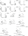
Measures and main results: We find that during IgG-IC-induced acute lung inflammation, PPARγ expression at both RNA and protein levels is repressed, which is consistent with the results obtained from macrophages treated with IgG-IC. Furthermore, both in vivo and in vitro data show that PPARγ activation reduces IgG-IC-mediated pro-inflammatory mediators' production, thereby alleviating lung injury. In terms of mechanism, we observe that the generation of Egr-1 elicited by IgG-IC is inhibited by PPARγ. As an important transcription factor, Egr-1 transcription is substantially increased by IgG-IC in both in vivo and in vitro studies, leading to augmented protein expression, thus amplifying IgG-IC-triggered expressions of inflammatory factors via association with their promoters.
Conclusion: During IgG-IC-stimulated acute lung inflammation, PPARγ activation can relieve the inflammatory response by suppressing the expression of its downstream target Egr-1 that directly binds to the promoter regions of several inflammation-associated genes. Therefore, regulation of PPARγ-Egr-1-pro-inflammatory mediators axis by PPARγ agonist Rosiglitazone may represent a novel strategy for blockade of acute lung injury.
Keywords: Egr-1; PPARγ; acute lung injury; inflammation; pro-inflammatory mediators.
Copyright © 2021 Yan, Chen, Ding, Zhou, Li, Deng, Yuan, Zhang and Wang.
Conflict of interest statement
The authors declare that the research was conducted in the absence of any commercial or financial relationships that could be construed as a potential conflict of interest.
Figures


Similar articles
-
Wogonin prevents lipopolysaccharide-induced acute lung injury and inflammation in mice via peroxisome proliferator-activated receptor gamma-mediated attenuation of the nuclear factor-kappaB pathway.Immunology. 2014 Oct;143(2):241-57. doi: 10.1111/imm.12305. Immunology. 2014. PMID: 24766487 Free PMC article.
-
Rosiglitazone promotes ENaC-mediated alveolar fluid clearance in acute lung injury through the PPARγ/SGK1 signaling pathway.Cell Mol Biol Lett. 2019 May 28;24:35. doi: 10.1186/s11658-019-0154-0. eCollection 2019. Cell Mol Biol Lett. 2019. PMID: 31160894 Free PMC article.
-
The protective effect of PPARγ in sepsis-induced acute lung injury via inhibiting PTEN/β-catenin pathway.Biosci Rep. 2020 May 29;40(5):BSR20192639. doi: 10.1042/BSR20192639. Biosci Rep. 2020. Retraction in: Biosci Rep. 2021 Apr 30;41(4):BSR-20192639_RET. doi: 10.1042/BSR-20192639_RET. PMID: 32420586 Free PMC article. Retracted.
-
Development, validation and implementation of an in vitro model for the study of metabolic and immune function in normal and inflamed human colonic epithelium.Dan Med J. 2015 Jan;62(1):B4973. Dan Med J. 2015. PMID: 25557335 Review.
-
Roles of Peroxisome Proliferator-Activated Receptor Gamma on Brain and Peripheral Inflammation.Cell Mol Neurobiol. 2018 Jan;38(1):121-132. doi: 10.1007/s10571-017-0554-5. Epub 2017 Oct 3. Cell Mol Neurobiol. 2018. PMID: 28975471 Free PMC article. Review.
Cited by
-
Protective effect of liriodendrin on IgG immune complex-induced acute lung injury via inhibiting SRC/STAT3/MAPK signaling pathway: a network pharmacology research.Naunyn Schmiedebergs Arch Pharmacol. 2023 Nov;396(11):3269-3283. doi: 10.1007/s00210-023-02534-1. Epub 2023 May 27. Naunyn Schmiedebergs Arch Pharmacol. 2023. PMID: 37243760
-
Calycosin attenuates renal ischemia/reperfusion injury by suppressing NF-κB mediated inflammation via PPARγ/EGR1 pathway.Front Pharmacol. 2022 Oct 7;13:970616. doi: 10.3389/fphar.2022.970616. eCollection 2022. Front Pharmacol. 2022. PMID: 36278223 Free PMC article.
-
Down-regulation of microRNA-155 suppressed Candida albicans induced acute lung injury by activating SOCS1 and inhibiting inflammation response.J Microbiol. 2022 Apr;60(4):402-410. doi: 10.1007/s12275-022-1663-5. Epub 2022 Feb 14. J Microbiol. 2022. PMID: 35157222 Free PMC article.
-
Early Growth Response-1, an Integrative Sensor in Cardiovascular and Inflammatory Disease.J Am Heart Assoc. 2021 Nov 16;10(22):e023539. doi: 10.1161/JAHA.121.023539. Epub 2021 Nov 10. J Am Heart Assoc. 2021. PMID: 34755520 Free PMC article. Review.
-
Single-cell transcriptomics unveiled that early life BDE-99 exposure reprogrammed the gut-liver axis to promote a proinflammatory metabolic signature in male mice at late adulthood.Toxicol Sci. 2024 Jun 26;200(1):114-136. doi: 10.1093/toxsci/kfae047. Toxicol Sci. 2024. PMID: 38648751 Free PMC article.
References
-
- Zubler RH, Nydegger U, Perrin LH, Fehr K, McCormick J, Lambert PH, et al. . Circulating and intra-articular immune complexes in patients with rheumatoid arthritis. Correlation of 125I-Clq binding activity with clinical and biological features of the disease. J Clin Invest (1976) 57:1308–19. 10.1172/JCI108399 - DOI - PMC - PubMed
Publication types
MeSH terms
Substances
LinkOut - more resources
Full Text Sources
Other Literature Sources
Research Materials

