

Acute appendiceal diverticulitis: a case report
- PMID: 33717385
- PMCID: PMC7921191
- DOI: 10.1016/j.radcr.2021.02.034
Acute appendiceal diverticulitis: a case report
Erratum in
-
Erratum regarding missing patient consent statements in previously published articles.Radiol Case Rep. 2023 Jan 25;18(4):1641-1642. doi: 10.1016/j.radcr.2023.01.013. eCollection 2023 Apr. Radiol Case Rep. 2023. PMID: 36895595 Free PMC article.
Abstract
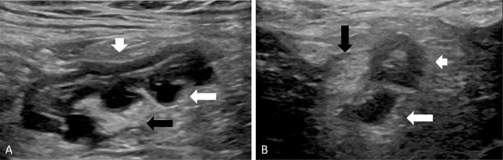
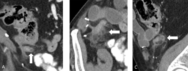
Acute appendiceal diverticulitis is a difficult prospective diagnosis due to both its rarity and clinical overlap with other ileocecal diseases, such as acute appendicitis. Few preoperative radiological images of acute appendiceal diverticulitis have been presented in the literature. We report a case of acute appendiceal diverticulitis initially diagnosed with outpatient ultrasound and confirmed with contrast-enhanced computed tomography in an 85-year-old male who presented with right lower quadrant pain. Appendiceal diverticulitis is an often overlooked diagnosis, and given its association with appendiceal neoplasms and relatively high rate of perforation, it is important to consider this entity in patients with right lower quadrant pain in order to ensure that they are managed appropriately. The addition of high quality ultrasound and computed tomography (CT) images to the literature may heighten awareness of this rare condition and facilitate accurate preoperative diagnosis and management in these patients.
Keywords: Abdominal pain; Appendiceal diverticulitis; Appendicitis; Ultrasound.
© 2021 The Authors.
Figures
References
-
- Kabiri H, Clarke LE, Tzarnas CD. Appendiceal diverticulitis. Am Surg. 2006;72(3):221–223. - PubMed
-
- Seker D, Seker G, Kahramanca S, Gurler M, Turker A, Kulacoglu H. A rare but distinctive cause of acute abdomen: appendiceal diverticulitis. J Emerg Med. 2013;44(1):e61–e62. - PubMed
-
- Yamana I, Kawamoto S, Inada K, Nagao S, Yoshida T, Yamashita Y. Clinical characteristics of 12 cases of appendiceal diverticulitis: a comparison with 378 cases of acute appendicitis. Surg Today. 2012;42(4):363–367. - PubMed
Publication types
LinkOut - more resources
Full Text Sources
Other Literature Sources
