. 2021 Feb;13(2):1011-1019.
doi: 10.21037/jtd-20-3032.

Imaging and monitoring in minimally invasive valve surgery using an intra-aortic occlusion device: a single center experience

Affiliations

Imaging and monitoring in minimally invasive valve surgery using an intra-aortic occlusion device: a single center experience

Calogera Pisano et al. J Thorac Dis. 2021 Feb.

Abstract

Background: Minimally invasive approach through a right mini-thoracotomy is a world-wide used procedure for mitral valve surgery. We performed a retrospective analysis based on our center experience in order to propose an effective, safe and reproducible method using an intra-aortic occlusion device.

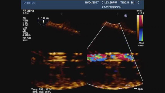
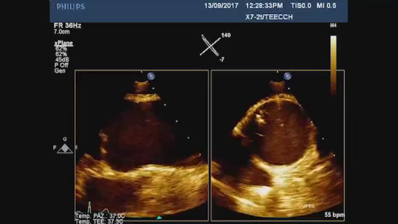
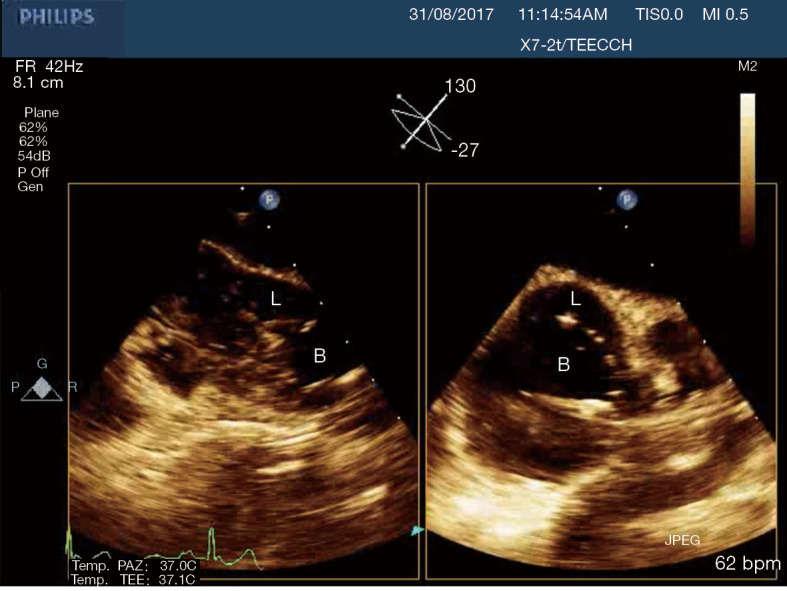
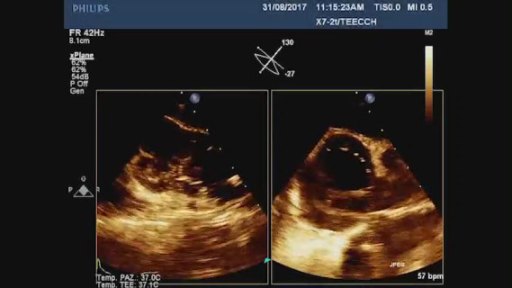
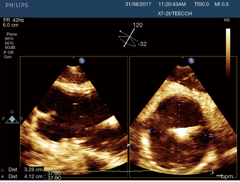
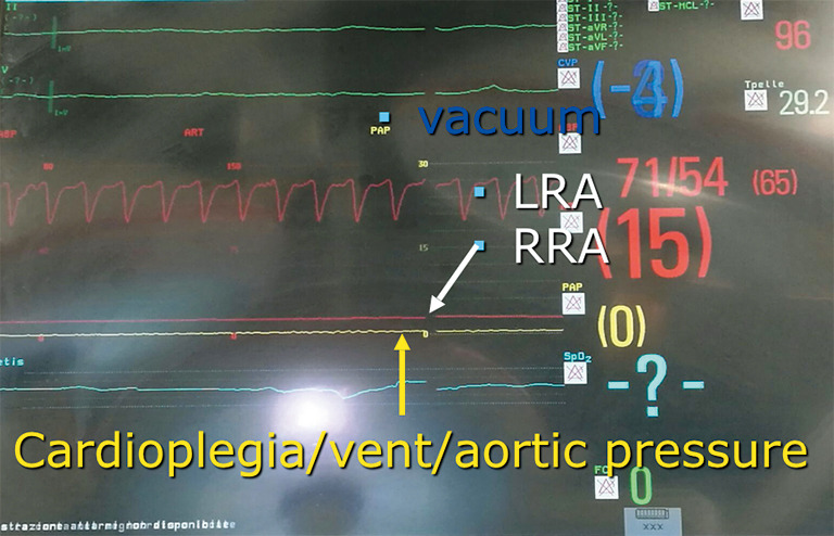
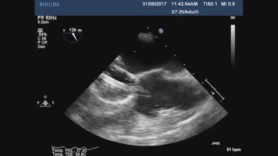
Methods: This is a retrospective analysis on 48 consecutive patients undergoing mitral valve surgery through a right anterolateral mini-thoracotomy in our center. An intra-aortic occlusion device was used for aortic clamping and cardioplegia delivery. Simultaneous multi-plane three-dimensional echocardiography imaging was acquired to detect the venous cannulas position, the intra-aortic device location in the ascending aorta, the balloon inflation, the complete occlusion of the aorta, the cardioplegia delivery, the origin and the blood flow in the right coronary artery. Aortic root pressure was measured by the tip of the intra-aortic occlusion device. A bilateral upper extremity invasive arterial pressure monitoring was detected. Neuromonitoring was performed through bilateral cerebral oximetry.

Results: The analysis has shown no aortic dissection, neurological damage type 1 and myocardial ischemia in the study population. In 3 cases a distal displacement of the intra-aortic occlusion device was promptly detected by the combined use of echocardiographic imaging and by a drop of the right cerebral oximetry saturation and of the right radial artery pressure.

Conclusions: The combined use of transesophageal simultaneous multi-plane three- dimensional echocardiography imaging, bilateral upper extremity invasive arterial pressure monitoring, aortic root pressure and cerebral oximetry is an effective, safe and reproducible method in patients undergoing minimally invasive valve surgery using an intra-aortic occlusion device.

Keywords: Minimally invasive approach; intra-aortic occlusion device; mitral valve surgery; three dimensional echocardiography.


Conflict of interest statement

Conflicts of Interest: All authors have completed the ICMJE uniform disclosure form (available at http://dx.doi.org/10.21037/jtd-20-3032). The authors have no conflicts of interest to declare.

Figures

Figure 1
Figure 1
TEE mid-esophageal bicaval view. Two stage cannulas passing through the right atrium. TEE, transesophageal echocardiography; IVC, inferior vena cava; SVC, superior vena cava; RA, right atrium.
Video 1
Video 1
TEE Midesophageal bicaval view. Wire in the correct position passing through the right atrium and entering in the superior vena cava.
Video 2
Video 2
TEE Midesophageal bicaval view dual color of a two stage venous cannula in a correct position during systemic perfusion.
Figure 2
Figure 2
Transesophageal identification of the right coronary sinus. Long axis view of high right coronary artery (RCA)’s origin (A). Short axis view of the coronaries origin (B).
Video 3
Video 3
TEE Simultaneous Multiplane Image of the ascending aorta. While the lumen of the aorta seems to be empty in a long axis view, the wire of the aortic occlusion device can be clearly detected in a short axis x-plane image.
Figure 3
Figure 3
Simultaneous multiplane transesophageal imaging of the ascending aorta. L, lumen; B, balloon.
Video 4
Video 4
TEE Simultaneous Multiplane image of the ascending aorta that shows an incomplete clamping of the vessel. Orthogonal short axis view that shows how the balloon doesn’t occupy the entire lumen of the aorta.
Figure 4
Figure 4
Simultaneous multiplane transesophageal echocardiography imaging of the ascending aorta. Long axis (A). Short axis (B). The diameters show an irregular shape of the vessel.
Figure 5
Figure 5
Hemodynamic monitoring during the procedure. Red Waves, bilateral invasive and continuous monitoring of the radial arteries. Yellow wave, aortic root pressure/Cardioplegia.
Video 5
Video 5
TEE Midesophageal long axis view: the increased pressure in the aortic root pushes the balloon distally towards the brachiocefalic trunk.
Figure 6
Figure 6
Right drop at the Cerebral Oximetry could indicate a distal migration of the balloon with an occlusion of the brachiocephalic trunk.
Figure 7
Figure 7
Distal displacement of the EAB. The occlusion of the brachiocephalic trunk determined a drop in the right radial artery pressure.
Figure 8
Figure 8
Identification of proximal displacement of the intra-aortic occlusion device. The systemic perfusion pressure exceeds aortic root pressure.
Video 6
Video 6
TEE Midesophageal long axis view: proxymal displacement of the intra aortic occlusion device. The balloon migrates into the left ventricle through the aortic valve.

References

    1. Murphy DA, Miller JS, Langford DA, et al. Endoscopic robotic mitral valve surgery. J Thorac Cardiovasc Surg 2006;132:776-81. 10.1016/j.jtcvs.2006.04.052 - DOI - PubMed
    1. Kara KA, Caner T. Comparison of pain in the early post-operative period using VAS score in patients after cardiac surgery who had minimally invasive incisions vs. full median sternotomy. Ann Ital Chir 2019;90:3-9. - PubMed
    1. Casselman FP, Van Slycke S, Wellens F, et al. Mitral valve surgery can now routinely be performed endoscop- ically. Circulation 2003;108:II48-54. 10.1161/01.cir.0000087391.49121.ce - DOI - PubMed
    1. Irace FG, Rose D, D'Ascoli R, et al. Video assistance in mitral surgery: reaching the "Thru" port access. J Vis Surg 2015;1:13. - PMC - PubMed
    1. Bentala M, Heuts S, Vos R, et al. Comparing the endo-aortic balloon and the external aortic clamp in minimally invasive mitral valve surgery. Interact CardioVasc Thorac Surg 2015;21:359-65. 10.1093/icvts/ivv160 - DOI - PubMed

LinkOut - more resources