. 2021 Feb 10:23:1288-1303.
doi: 10.1016/j.omtn.2021.02.003. eCollection 2021 Mar 5.

Tumor suppressor lnc-CTSLP4 inhibits EMT and metastasis of gastric cancer by attenuating HNRNPAB-dependent Snail transcription

Affiliations

Tumor suppressor lnc-CTSLP4 inhibits EMT and metastasis of gastric cancer by attenuating HNRNPAB-dependent Snail transcription

Tao Pan et al. Mol Ther Nucleic Acids. .

Abstract

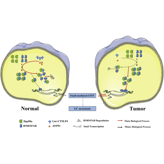
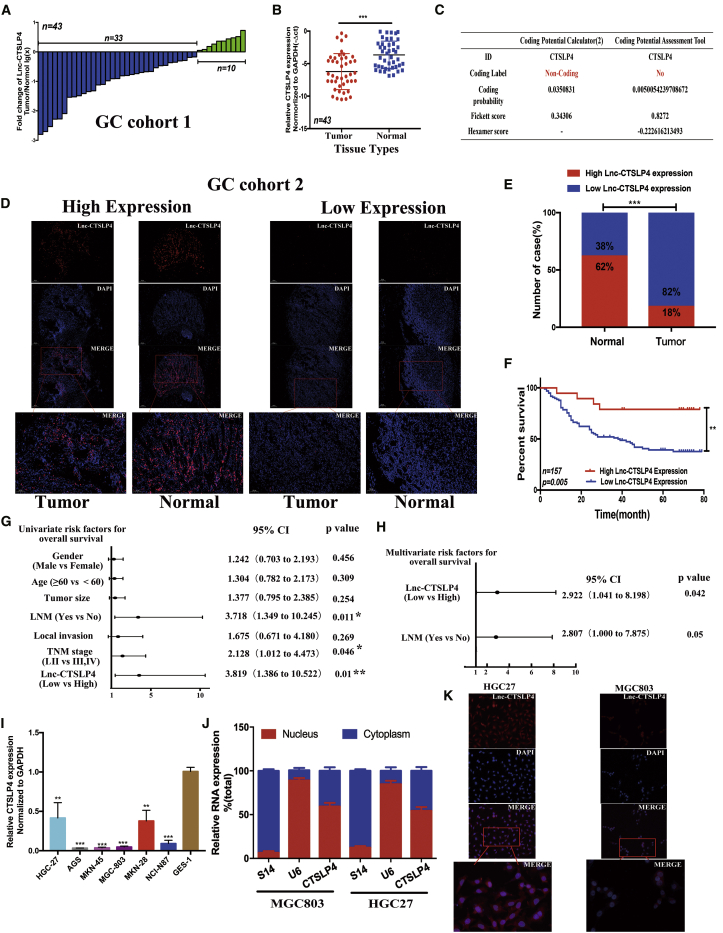
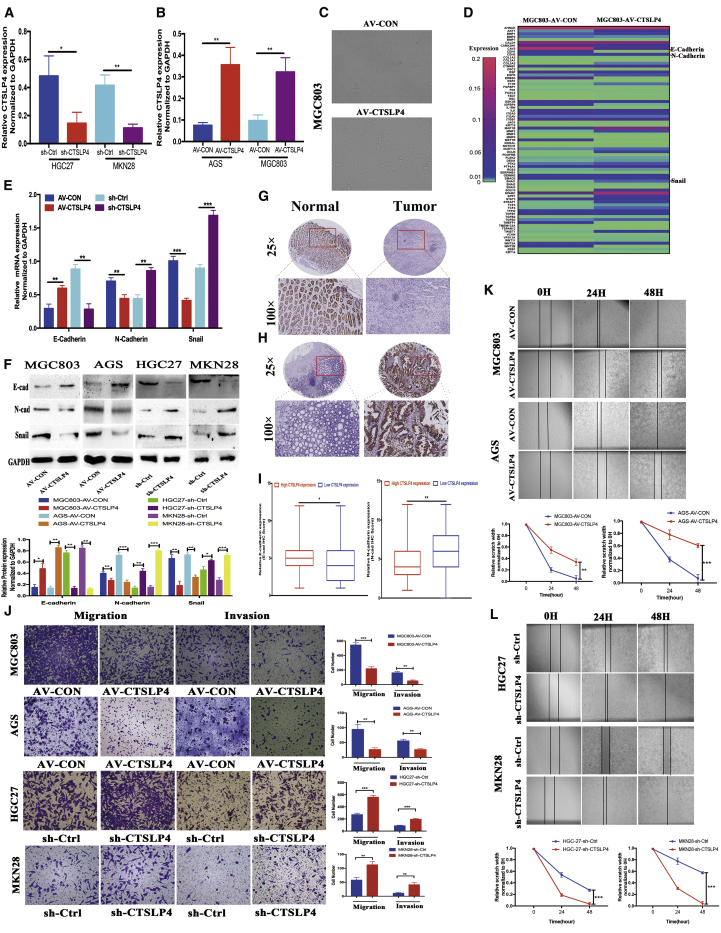
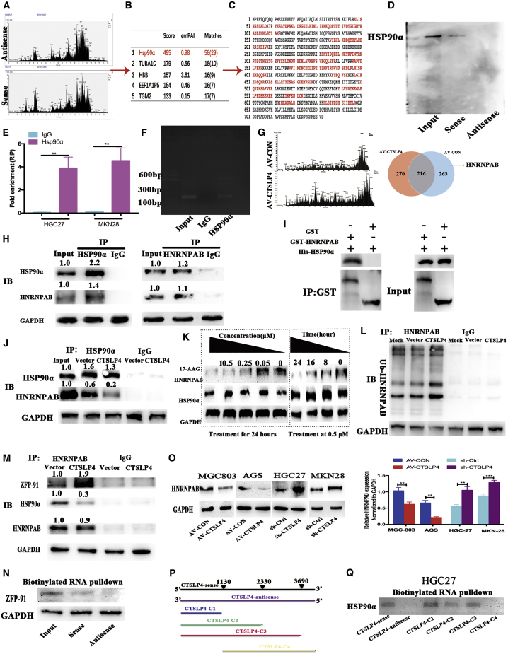
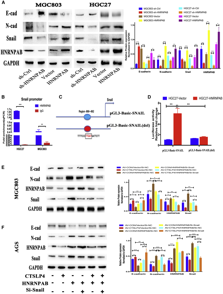
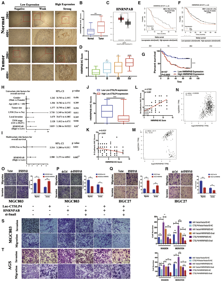
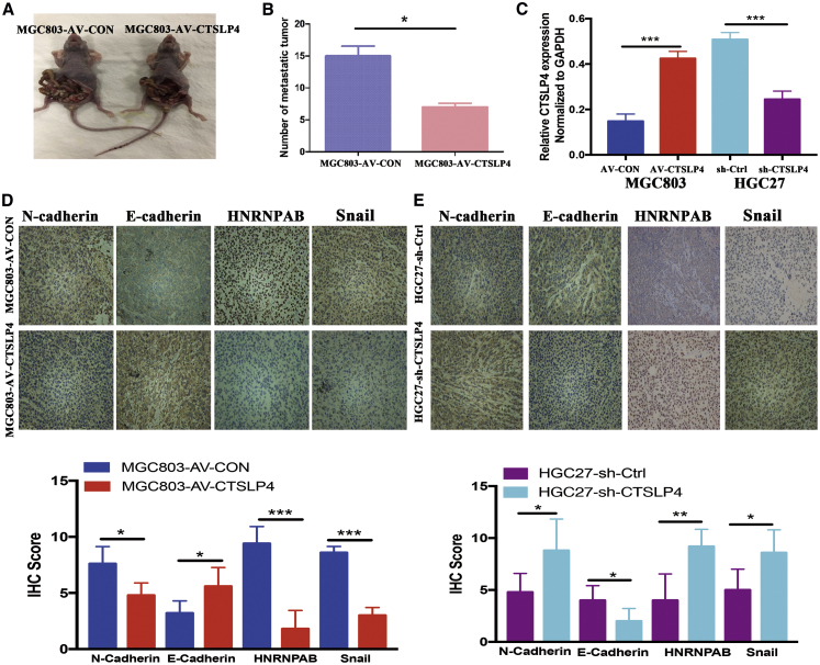
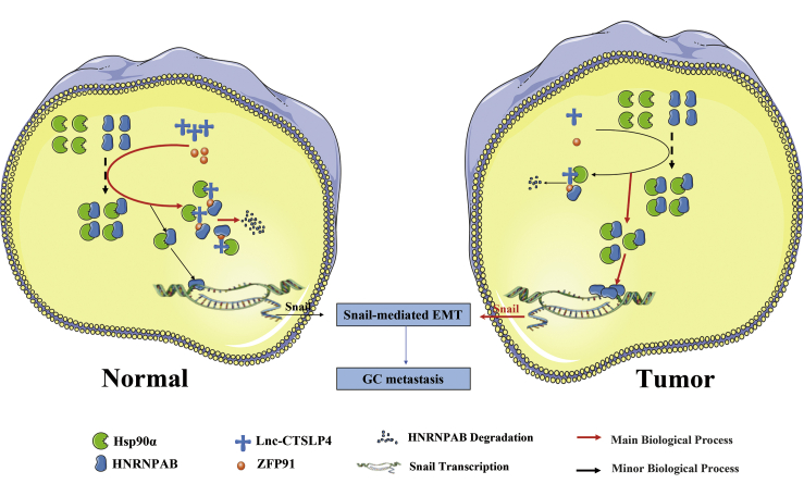
Tumor metastasis is a crucial impediment to the treatment of gastric cancer (GC), and the epithelial-to-mesenchymal transition (EMT) program plays a critical role for the initiation of GC metastasis. Thus, the aim of this study is to investigate the regulation of lnc-CTSLP4 in the EMT process during GC progression. We found that lnc-CTSLP4 was significantly downregulated in GC tumor tissues compared with adjacent non-tumor tissues, and its levels in GC tumor tissues were closely correlated with tumor local invasion, TNM stage, lymph node metastasis, and prognosis of GC patients. Loss- and gain-of-function assays indicated that lnc-CTSLP4 inhibited GC cell migration, invasion, and EMT in vitro, as well as peritoneal dissemination in vivo. Mechanistic analysis demonstrated that lnc-CTSLP4 could bind with Hsp90α/heterogeneous nuclear ribonucleoprotein AB (HNRNPAB) complex and recruit E3-ubiquitin ligase ZFP91 to induce the degradation of HNRNPAB, thus suppressing the transcriptional activation of Snail and ultimately reversing EMT of GC cells. Taken together, our results suggest that lnc-CTSLP4 is significantly downregulated in GC tumor tissues and inhibits metastatic potential of GC cells by attenuating HNRNPAB-dependent Snail transcription via interacting with Hsp90α and recruiting E3 ubiquitin ligase ZFP91, which shows that lnc-CTSLP4 could serve as a prognostic biomarker and therapeutic target for metastatic GC.

Keywords: EMT; Gastric Cancer; HNRNPAB; Hsp90α; Metastasis; Snail;LncRNA; Ubiquitin; ZFP91; lnc-CTSLP4.


Conflict of interest statement

The authors declare no competing interests.

Figures

None
Graphical abstract
Figure 1
Figure 1
Lnc-CTSLP4 expression is downregulated in tumor tissues and inversely associated with prognosis of GC patients (A) The expression of lnc-CTSLP4 in 43 GC tissues (GC cohort 1, the ratio of GC tumor tissues versus adjacent non-tumor tissues [−ΔCt]). (B) Expression levels of lnc-CTSLP4 in GC tumor tissues (GC cohort 1) and adjacent normal tissues (−ΔCt, ∗p < 0.05, ∗∗p < 0.01, ∗∗∗p < 0.001). (C) The coding ability of lnc-CTSLP4 calculated by CPC2 and CPAT. (D) RNA-FISH analysis of lnc-CTSLP4 expression in the paraffin-embedded GC tumor tissues and adjacent non-tumor tissues (red: lnc-CTSLP4; blue: nuclear; n = 157, scale bars: 100 μm and 200 μm). (E) lnc-CTSLP4 high and low expression rate in GC (GC cohort 2, RNA-FISH). (F) Kaplan-Meier analysis of the overall survival of GC (GC cohort 2, n = 157). The log-rank test revealed statistical significance between the low CTSLP4 expression group (n = 129) and the high CTSLP4 expression group (n = 28). (G and H) Univariate (G) and multivariate (H) analysis for prognostic features of GC patients (n = 157). (I) Expression of lnc-CTSLP4 in GC cell lines and a normal gastric epithelium cell line, GES-1. (J) The expression of lnc-CTSLP4 in the subcellular fractions of GC cells (MGC803 and HGC27, qRT-PCR; U6 and S14 were used as nuclear and cytoplasmic markers, respectively). (K) Representative RNA-FISH imaging of lnc-CTSLP4 (red) in GC cells (red: lnc-CTSLP4; blue: DAPI; magnification: 200× and 400×).
Figure 2
Figure 2
Lnc-CTSLP4 overexpression inhibits GC cell EMT, migration and invasion in vitro (A and B) qRT-PCR analysis of lnc-CTSLP4 expression in GC cells after shRNA knockdown (A) or overexpression (B) of lnc-CTSLP4. ∗p < 0.05, ∗∗p < 0.01. (C) Morphological change of MGC803 cells after overexpression of CTSLP4 (100×). (D) PCR-array analysis of EMT-associated genes in MGC803-AV-CTSLP4 and MGC803-AV-CON cells. (E) qRT-PCR analysis of E-cadherin, N-cadherin, and Snail mRNA in GC cells after overexpression or shRNA knockdown of lnc-CTSLP4. ∗∗p < 0.01, ∗∗∗p < 0.001. (F) Western blot analysis of E-cadherin, N-cadherin, and Snail protein in GC cells (MGC803, AGS, HGC27, MKN28 cells) after knockdown of lnc-CTSLP4 (∗p < 0.05, ∗∗p < 0.01, ∗∗∗p < 0.001). (G and H) Representative IHC imaging of E-cadherin and N-cadherin (magnification: 25× and 100×). (I) Correlation of lnc-CTSLP4 expression (RNA-FISH) and EMT makers (E-cadherin and N-cadherin, IHC) in GC (GC cohort 2, ∗p < 0.05, ∗∗p < 0.01). (J) The migration and invasion abilities of GC cells after overexpression or knockdown of lnc-CTSLP4 (Transwell assay, ∗∗p < 0.01, ∗∗∗p < 0.001, 200×). (K and L) Wound-healing assays of GC cells after transfection with AV-CTSLP4 or sh-CTSLP4 (∗∗p < 0.01, ∗∗∗p < 0.001, magnification: 200×).
Figure 3
Figure 3
Lnc-CTSLP4 binds to Hsp90α and promotes the degradation of HNRNPAB protein by recruiting ZFP91 (A and B) RNA-pulldown assays and LC-MS/MS analysis of proteins that are specifically bound to lnc-CTSLP4 (anti-sense RNA as a negative control, A represents proteins that bound to sense or antisense of lnc-CTSLP4 and B represents top 5 proteins identified). (C) The peptide sequences of Hsp90α binding with lnc-CTSLP4 (red: matched peptide sequences). (D) Hsp90α was detected by Western blot after RNA-pulldown assays (A). (E and F) RIP and qRT-PCR analysis for the specific binding of Hsp90α to lnc-CTSLP4. ∗∗p < 0.01. (G) Co-IP followed by LC-MS/MS analysis showed that the Hsp90α could bind to HNRNPAB in the absence of lnc-CTSLP4. (H) The interaction of Hsp90α and HNRNPAB was verified by Co-IP in MGC803 cells. (I) GST-pulldown analysis of the binding of Hsp90α and HNRNPAB. (J) Analysis of proteins obtained in Co-IP in lnc-MGC803-AV-CTSLP4 and MGC803-AV-CON cells. (K) The protein levels of HNRNPAB and Hsp90α in MGC803 cells treated with 17-AAG (an Hsp90 inhibitor) for the indicated doses and times. (L) Ubiquitin modification of HNRNPAB in MGC803-AV-CTSLP4 and MGC803-AV-CON cells. (M) Co-IP and Western blot analysis of the binding of HNRNPAB to ZFP91 or Hsp90α in MGC803-AV-CTSLP4 and MGC803-AV-CON cells. (N) The specific binding of ZFP91 to lnc-CTSLP4 verified by RNA-pulldown assays (negative control: anti-sense). (O) Western blot analysis of HNRNPAB protein in GC cells (MGC803, AGS, HGC27, MKN28) after overexpression (AV-CTSLP4) or shRNA knockdown of lnc-CTSLP4 (sh-CTSLP4). (P) Truncated versions of lnc-CTSLP4, C1 (1-1130 bp), C2 (1-2330 bp), C3 (1-3690 bp), and C4 (1130-3690 bp) according to the predicted lnc-CTSLP4 structure. (Q) Western blot detection of Hsp90α after RNA-pulldown with biotinylated RNAs for different constructs of lnc-CTSLP4 or its antisense strand (negative control).
Figure 4
Figure 4
Lnc-CTSLP4 inhibits EMT via suppressing HNRNPAB-mediated Snail transcriptional activation in GC cells (A) The expression level of E-cadherin, N-cadherin, HNRNPAB, and Snail in MGC803 and HGC27 cells after shRNA knockdown (sh-HNRNPAB) or overexpression (HNRNPAB) of HNRNPAB (∗∗p < 0.01, ∗∗∗p < 0.001). (B) The binding of HNRNPAB to the Snail promoter in GC cells (ChIP-PCR assays, ∗∗p < 0.01, ∗∗∗p < 0.001). (C and D) pGL3-Basic-SNAIL and pGL3-Basic-SNAIL-del (deletion at position −866 to −862 bp) constructs, along with the pRL-TK plasmid, were transfected into HGC27-HNRNPAB cells and HGC27-Vector cells, and then the relative luciferase activity was measured (∗∗p < 0.01, ∗∗∗p < 0.001). (E and F) Western blot (E) and densitometric analysis (F) of E-cadherin, N-cadherin, HNRNPAB, and Snail in MGC803 and AGS cells transfected with AV-CTSLP4, HNRNPAB-expressing vector, or Snail si-RNA (∗∗p < 0.01, ∗∗∗p < 0.001).
Figure 5
Figure 5
HNRNPAB is inversely correlated with lnc-CTSLP4 in GC tissues and rescues the suppressive effects of lnc-CTSLP4 on GC cells (A) Representative immunohistochemical staining of HNRNPAB in GC tumor tissues and adjacent non-tumor tissues (magnification: 200× and 400×). (B) The expression levels of HNRNPAB in 157 pairs of GC patients based on HNRNPAB IHC score (GC cohort 2, ∗∗∗p < 0.001). (C) Validation of HNRNPAB expression in GEPIA. (D) The expression levels of HNRNPAB in GC patients with different pathological stages based on HNRNPAB IHC score (∗∗p < 0.01, ∗∗∗p < 0.001). (E and F) The correlation of HNRNPAB expression levels and overall survival in the GSE14210 dataset (n = 118) and GSE14210, GSE15459, GSE22377, GSE29272, and GSE51105 dataset (n = 592) was analyzed by Kaplan-Meier plotter. (G) Kaplan-Meier survival analysis for OS of 157 patients with GC (GC cohort 2) reveals a statistically significant difference between the low HNRNPAB expression group (n = 129) and the high HNRNPAB group (n = 28). (H and I) Univariate (H) and multivariate (I) analysis of for prognostic features of GC patients (n = 157) showed that the expression of HNRNPAB was an independent prognostic factor for survival. (J) HNRNPAB is downregulated in the high lnc-CTSLP4 expression group compared to the low lnc-CTSLP4 expression group (GC cohort 2, ∗∗∗p < 0.001). (K and L) Correlation analysis of HNRNPAB with E-cadherin (K) or N-cadherin (L) in GC. (M and N) Validation of the relationship between HNRNPAB and E-cadherin (M) or Snail (N) based on GEPIA. (O–R) The metastatic abilities of GC cells (MGC803 and HGC27) after overexpression or shRNA knockdown of HNRNPAB (Transwell assays, magnification: 200×). (S and T) The metastatic abilities of GC cells (MGC803 and AGS cells) transfected with lnc-CTSLP4, HNRNPAB-expressing vector, or Snail si-RNA (Transwell assays, magnification: 200×, ∗∗∗p < 0.001).
Figure 6
Figure 6
Lnc-CTSLP4 overexpression inhibits GC cell invasion and metastasis in vivo (A) The effect of CTSLP4 overexpression in MGC803 cell-derived peritoneal nodules in nude mice (n = 5 per group, ∗p < 0.05). (B) Average numbers of peritoneal nodules from nude mice. (C) The levels of lnc-CTSLP4 expression in tumor tissues formed from MGC803 cells (transfected with AV-CTSLP4 or AV-CON) and HGC27 cells (stably transfected with sh-CTSLP4 or sh-Ctrl lentivirus, ∗∗∗p < 0.001). (D) Representative immunohistochemical staining for E-cadherin, N-cadherin, HNRNPAB, and Snail in xenograft tumors derived from MGC803 cells transfected with AV-CTSLP4 or AV-CON (n = 5 per group, magnification: 200×). (E) Representative immunohistochemical staining for E-cadherin, N-cadherin, HNRNPAB, and Snail in xenograft tumors derived from HGC27 cells transfected with sh-CTSLP4 or sh-Ctrl lentivirus (n = 5 per group, magnification: 200×, ∗p < 0.05).
Figure 7
Figure 7
Schematic of the proposed mechanism of lnc-CTSLP4 in GC progression Lnc-CTSLP4, as a regulator with decreased levels in GC, binds to Hsp90α and recruits the E3 ubiquitin ligase ZFP91, which can block the interaction of Hsp90α with HNRNPAB and decreases the stability of HNRNPAB, subsequently promotes its degradation, thus reducing the transcriptional activation of Snail and inhibiting Snail-mediated EMT.

Similar articles

Cited by

References

    1. Bray F., Ferlay J., Soerjomataram I., Siegel R.L., Torre L.A., Jemal A. Global cancer statistics 2018: GLOBOCAN estimates of incidence and mortality worldwide for 36 cancers in 185 countries. CA Cancer J. Clin. 2018;68:394–424. - PubMed
    1. Chen W., Zheng R., Baade P.D., Zhang S., Zeng H., Bray F., Jemal A., Yu X.Q., He J. Cancer statistics in China, 2015. CA Cancer J. Clin. 2016;66:115–132. - PubMed
    1. Bernards N., Creemers G.J., Nieuwenhuijzen G.A., Bosscha K., Pruijt J.F., Lemmens V.E. No improvement in median survival for patients with metastatic gastric cancer despite increased use of chemotherapy. Ann. Oncol. 2013;24:3056–3060. - PubMed
    1. Wagner A.D., Syn N.L., Moehler M., Grothe W., Yong W.P., Tai B.C., Ho J., Unverzagt S. Chemotherapy for advanced gastric cancer. Cochrane Database Syst. Rev. 2017;8:CD004064. - PMC - PubMed
    1. Chaffer C.L., Weinberg R.A. A perspective on cancer cell metastasis. Science. 2011;331:1559–1564. - PubMed