Review
. 2021;8(3):167-177.
doi: 10.1007/s40588-021-00160-0. Epub 2021 Mar 5.

Cardiovascular Injury Due to SARS-CoV-2

Affiliations
Review

Cardiovascular Injury Due to SARS-CoV-2

Christina L Bugert et al. Curr Clin Microbiol Rep. 2021.

Abstract

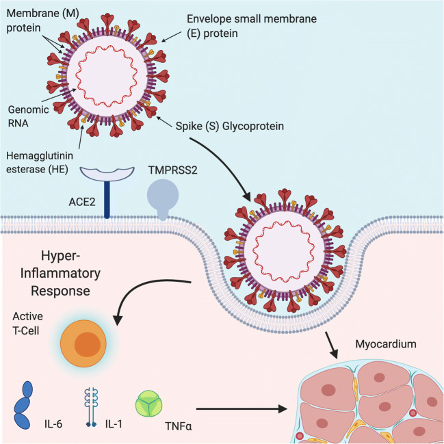
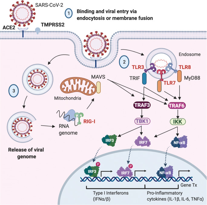
Purpose of review: The world is currently facing the largest global health crisis since the early 1900s due to a novel coronavirus. While SARS-CoV-2 infection causes predictable symptoms in COVID-19 patients, including upper respiratory distress and fever, the heterogeneity of manifestations is surprising. This review focuses on direct and indirect causes of myocardial injury in COVID-19 patients and highlights current knowledge, treatment strategies, and outstanding questions in the field.

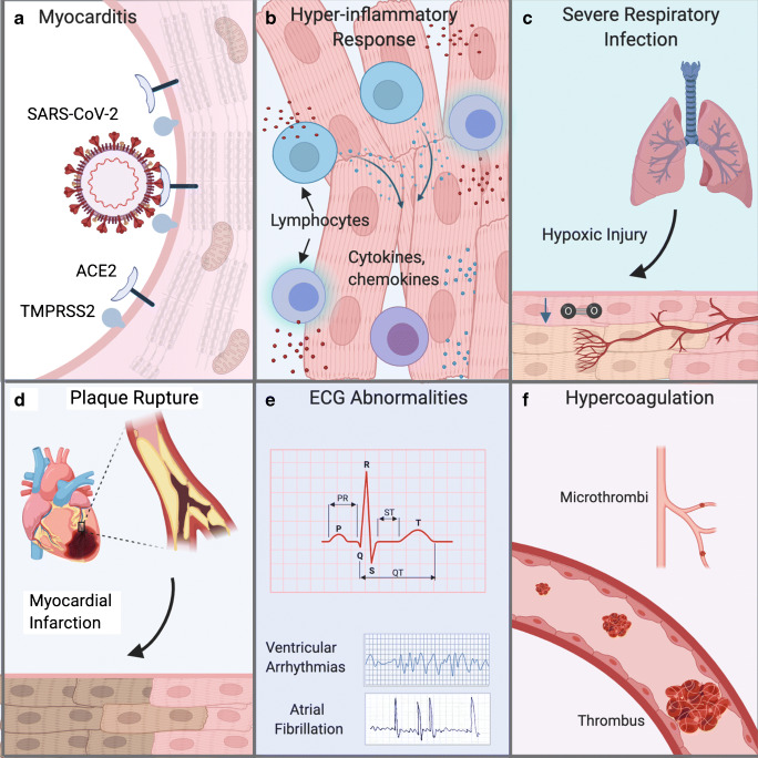
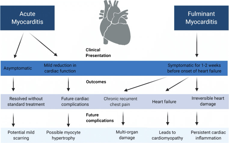
Recent findings: Data are emerging that highlight the extent of cardiovascular involvement in COVID-19 patients, including evidence that SARS-CoV-2 causes myocarditis and increases cardiac risk. The incidence of cardiac injury is much greater in patients with severe disease presentation and those in intensive care.

Summary: During the past year, COVID-19 patient mortality rates have improved due to tailored pharmacological treatments and patient management strategies that address the unique presentation of symptoms, which will hopefully also reduce the incidence of cardiac injury.

Keywords: Cardiac injury; Arrhythmia; COVID-19; Cardiomyopathy; Myocarditis.


Figures

Fig. 1
Fig. 1
Mechanism of SARS-CoV-2 Cellular. Entry into host cells is an important determinant of infectivity and infection and pathogenesis of viruses. SARS-CoV-2 has various proteins on its surface including (M) membrane protein, (E) envelope small membrane protein and (S) spike glycoproteins. Interaction of the SARS-CoV-2 spike protein with angiotensin-converting enzyme 2 (ACE2) receptors on target the cell surface mediates SARS-CoV-2 entry into cells that is facilitated by transmembrane protease serine 2 (TMPRSS2) and lysosomal proteases. Once SARS-CoV-2 enters the body it can elicit a hyper inflammatory response characterized by high levels of interleukin-6 (IL-6), interleukin-1 (IL-1), tumor necrosis factor-alpha (TNFα) that can damage the myocardium or through direct infection of cardiomyocytes which can damage the heart. Activated T-cells are one of the immune cells involved in the antiviral response. Figure created using BloRender.com
Fig. 2
Fig. 2
Various Routes to Cardiac Injury Due to SARS-CoV-2 Infection. (A) Several studies suggest that SARS-CoV-2 can directly infect cardiac cells of the myocardium and may cause heart damage by a “direct effect“. (B) The heart can also be injured by a systematic hyper inflammatory response that has been reported in COVID-19 patients by an “indirect effect“. High levels of cytokines and chemokines can cause ”bystander effects” and cardiac inflammation also classified as myocarditis or weakening of heart muscle function and may manifest as dilated cardiomyopathy (DCM) and/or heart failure. (C) Severe respiratory infection of SARS-CoV-2 infection can reduce pulmonary function and cause hypoxic injury in many organs including the heart due to low tissue oxygenation. (D) Increased stress on the cardiovascular system of COVID-19 patients can worsen underlying cardiac risk factors including rupture of atherosclerotic plaques leading to ST-segment myocardial infarction (STEMI) myocardial infarction. COVID-19 patients with severe infection can also exhibit non-STEMI myocardial infarction. (E) Various alterations in heart rhythm may occur as the first cardiac symptom in COVID-19 patients and can manifest as ventricular arrhytmias and atrial fibrillation that are detectable by electrocardiogram (ECG). (F) Coagulation abnormalities lead to formation of thrombi that can block blood flow in larger vessels or microthrombi that occlude small blood vessels and capillaries to affect microcirculation. The location and size of the blood vessel blockage dictate extent of ischemic tissue damage. Figure created with BioRender.com
Fig. 3
Fig. 3
Innate Immune Activation in SARS-CoV-2 Infection. The SARS-CoV-2 virus gains entry to cells by (1) binding to angiotensin converting enzyme 2 (ACE2) receptors on the cell surface through endocytosis or membrane fusion. (2) After entry the (+ sense) viral genome is released into the cytoplasm, (3) ribosomes (periwinkle) bind to viral genome to translate viral message into viral polymerase protein (light blue) and (4) RNA replication to produce (- sense) RNA genomes. A hyper inflammatory response can occur in SARS-CoV-2 infection and the presence of viral genomic and subgenomic RNA species activate cytosolic sensors such as toll-like receptor 3, 7 and 8 (TLR3/TLR7/TKR8). The activation of TRAF-6 leads to nuclear localization of NF-kB, IRF5 and IRF7 to induce expression of interferon stimulated genes that encode antiviral proteins and pro-inflammatory cytokines. Activation of TRAF-3 causes IRF3 to assume nuclear localization to stimulate production of Type I/II interferons. Produced using BioRender.com and adapted from a template provided by Natalya Odoardi Clinical Research Coordinator, Children’s Hospital of London
Fig. 4
Fig. 4
Myocarditis due to SARS-CoV-2 Infection. Myocarditis can manifest in two distinct presentations and cause acute or fulminant infection of heart tissue which can lead to future long-term complications

Similar articles

Cited by

References

    1. Chan JF, et al. A familial cluster of pneumonia associated with the 2019 novel coronavirus indicating person-to-person transmission: a study of a family cluster. Lancet. 2020;395(10223):514–523. doi: 10.1016/S0140-6736(20)30154-9. - DOI - PMC - PubMed
    1. Zhou P, Yang XL, Wang XG, Hu B, Zhang L, Zhang W, Si HR, Zhu Y, Li B, Huang CL, Chen HD, Chen J, Luo Y, Guo H, Jiang RD, Liu MQ, Chen Y, Shen XR, Wang X, Zheng XS, Zhao K, Chen QJ, Deng F, Liu LL, Yan B, Zhan FX, Wang YY, Xiao GF, Shi ZL. A pneumonia outbreak associated with a new coronavirus of probable bat origin. Nature. 2020;579(7798):270–273. - PMC - PubMed
    1. Nishiga M, Wang DW, Han Y, Lewis DB, Wu JC. COVID-19 and cardiovascular disease: from basic mechanisms to clinical perspectives. Nat Rev Cardiol. 2020;17(9):543–558. doi: 10.1038/s41569-020-0413-9. - DOI - PMC - PubMed
    1. Zhu N, Zhang D, Wang W, Li X, Yang B, Song J, Zhao X, Huang B, Shi W, Lu R, Niu P, Zhan F, Ma X, Wang D, Xu W, Wu G, Gao GF, Tan W, China Novel Coronavirus Investigating and Research Team A novel coronavirus from patients with pneumonia in China, 2019. N Engl J Med. 2020;382(8):727–733. doi: 10.1056/NEJMoa2001017. - DOI - PMC - PubMed
    1. Bavishi C, Maddox TM, Messerli FH. Coronavirus disease 2019 (COVID-19) infection and renin angiotensin system blockers. JAMA Cardiol. 2020;5(7):745–747. doi: 10.1001/jamacardio.2020.1282. - DOI - PubMed

LinkOut - more resources