

Comparison of the Gross Target Volumes Based on Diagnostic PET/CT for Primary Esophageal Cancer
- PMID: 33718127
- PMCID: PMC7947883
- DOI: 10.3389/fonc.2021.550100
Comparison of the Gross Target Volumes Based on Diagnostic PET/CT for Primary Esophageal Cancer
Abstract
Background: Clinically, many esophageal cancer patients who planned for radiation therapy have already undergone diagnostic Positron-emission tomography/computed tomography (PET/CT) imaging, but it remains unclear whether these imaging results can be used to delineate the gross target volume (GTV) of the primary tumor for thoracic esophageal cancer (EC).
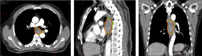
Methods: Seventy-two patients diagnosed with thoracic EC had undergone prior PET/CT for diagnosis and three-dimensional CT (3DCT) for simulation. The GTV3D was contoured on the 3DCT image without referencing the PET/CT image. The GTVPET-ref was contoured on the 3DCT image referencing the PET/CT image. The GTVPET-reg was contoured on the deformed registration image derived from 3DCT and PET/CT. Differences in the position, volume, length, conformity index (CI), and degree of inclusion (DI) among the target volumes were determined.
Results: The centroid distance in the three directions between two different GTVs showed no significant difference (P > 0.05). No significant difference was found among the groups in the tumor volume (P > 0.05). The median DI values of the GTVPET-reg and GTVPET-ref in the GTV3D were 0.82 and 0.86, respectively (P = 0.006). The median CI values of the GTV3D in the GTVPET-reg and GTVPET-ref were 0.68 and 0.72, respectively (P = 0.006).
Conclusions: PET/CT can be used to optimize the definition of the target volume in EC. However, no significant difference was found between the GTVs delineated based on visual referencing or deformable registration whether using the volume or position. So, in the absence of planning PET-CT images, it is also feasible to delineate the GTV of primary thoracic EC with reference to the diagnostic PET-CT image.
Keywords: 18F-FDG PET/CT; deformable image registration; gross target volume; thoracic esophageal cancer; three-dimensional computed tomography.
Copyright © 2021 Shi, Li, Li, Zhang, Guo, Wang and Wang.
Conflict of interest statement
The authors declare that the research was conducted in the absence of any commercial or financial relationships that could be construed as a potential conflict of interest.
Figures

Similar articles
-
Geometrical differences in target volumes based on 18F-fluorodeoxyglucose positron emission tomography/computed tomography and four-dimensional computed tomography maximum intensity projection images of primary thoracic esophageal cancer.Dis Esophagus. 2014 Nov-Dec;27(8):744-50. doi: 10.1111/dote.12247. Epub 2014 Jun 11. Dis Esophagus. 2014. PMID: 24915760
-
A comparative study of target volumes based on 18F-FDG PET-CT and ten phases of 4DCT for primary thoracic squamous esophageal cancer.Onco Targets Ther. 2017 Jan 6;10:177-184. doi: 10.2147/OTT.S95322. eCollection 2017. Onco Targets Ther. 2017. PMID: 28123302 Free PMC article.
-
Geometrical Comparison and Quantitative Evaluation of 18F-FDG PET/CT- and DW-MRI-Based Target Delineation Before and During Radiotherapy for Esophageal Squamous Carcinoma.Front Oncol. 2021 Dec 22;11:772428. doi: 10.3389/fonc.2021.772428. eCollection 2021. Front Oncol. 2021. PMID: 35004291 Free PMC article.
-
Comparison of primary target volumes delineated on four-dimensional CT and 18 F-FDG PET/CT of non-small-cell lung cancer.Radiat Oncol. 2014 Aug 15;9:182. doi: 10.1186/1748-717X-9-182. Radiat Oncol. 2014. PMID: 25123450 Free PMC article.
-
Comparison of gross target volumes based on four-dimensional CT, positron emission tomography-computed tomography, and magnetic resonance imaging in thoracic esophageal cancer.Cancer Med. 2020 Aug;9(15):5353-5361. doi: 10.1002/cam4.3072. Epub 2020 Jun 8. Cancer Med. 2020. PMID: 32510183 Free PMC article.
Cited by
-
The Role of MRI and PET/CT in Radiotherapy Target Volume Determination in Gastrointestinal Cancers-Review of the Literature.Cancers (Basel). 2023 May 29;15(11):2967. doi: 10.3390/cancers15112967. Cancers (Basel). 2023. PMID: 37296929 Free PMC article. Review.
-
Condition control training-based ConVMLP-ResU-Net for semantic segmentation of esophageal cancer in 18F-FDG PET/CT images.Phys Eng Sci Med. 2023 Dec;46(4):1643-1658. doi: 10.1007/s13246-023-01327-3. Epub 2023 Nov 1. Phys Eng Sci Med. 2023. PMID: 37910383
-
Gross Tumor Volume Definition and Comparative Assessment for Esophageal Squamous Cell Carcinoma From 3D 18F-FDG PET/CT by Deep Learning-Based Method.Front Oncol. 2022 Mar 17;12:799207. doi: 10.3389/fonc.2022.799207. eCollection 2022. Front Oncol. 2022. PMID: 35372054 Free PMC article.
-
Inter-observer variation in gross tumour volume delineation of oesophageal cancer on MR, CT and PET/CT.Radiol Oncol. 2024 Oct 4;58(4):580-587. doi: 10.2478/raon-2024-0043. eCollection 2024 Dec 1. Radiol Oncol. 2024. PMID: 39362222 Free PMC article.
References
-
- Zou B, Tu Y, Liao D, Xu Y, Wang J, Huang M, et al. . Radical esophagectomy for stage II and III thoracic esophageal squamous cell carcinoma followed by adjuvant radiotherapy with or without chemotherapy: Which is more beneficial? Thoracic Cancer (2020) 11(3):631–9. 10.1111/1759-7714.13307 - DOI - PMC - PubMed
-
- Ikeguchi M, Kohno Y, Kihara K, Suzuki K, Saito H. Neoadjuvant chemotherapy for clinical stage ii and iii thoracic esophageal squamous cell carcinoma with curative esophagectomy. J Cancer Ther (2015) 06(15):1207–13. 10.4236/jct.2015.615131 - DOI
LinkOut - more resources
Full Text Sources
Other Literature Sources