

Positron Emission Tomography-Based Short-Term Efficacy Evaluation and Prediction in Patients With Non-Small Cell Lung Cancer Treated With Hypo-Fractionated Radiotherapy
- PMID: 33718144
- PMCID: PMC7947869
- DOI: 10.3389/fonc.2021.590836
Positron Emission Tomography-Based Short-Term Efficacy Evaluation and Prediction in Patients With Non-Small Cell Lung Cancer Treated With Hypo-Fractionated Radiotherapy
Abstract
Background: Positron emission tomography is known to provide more accurate estimates than computed tomography when staging non-small cell lung cancer. The aims of this prospective study were to contrast the short-term efficacy of the two imaging methods while evaluating the effects of hypo-fractionated radiotherapy in non-small cell lung cancer, and to establish a short-term efficacy prediction model based on the radiomics features of positron emission tomography.
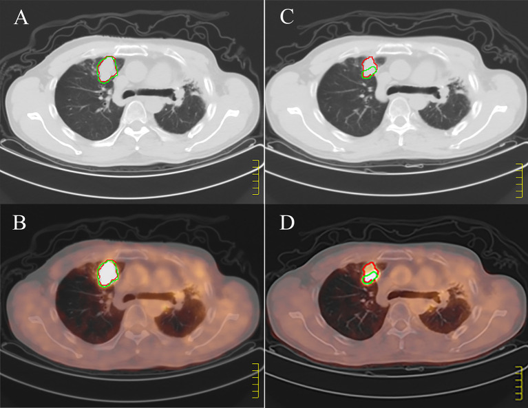
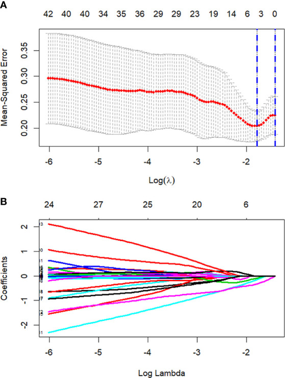
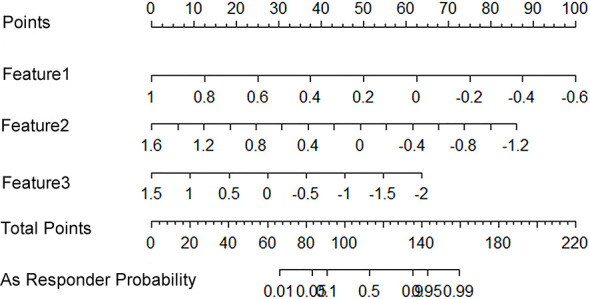
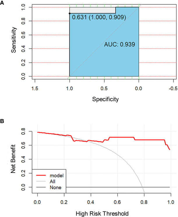
Methods: This nonrandomized-controlled trial was conducted from March 2015 to June 2019. Thirty-one lesions of 30 patients underwent the delineation of the regions of interest on positron emission tomography and computed tomography 1 month before, and 3 months after hypo-fractionated radiotherapy. Each patient was evaluated for the differences in local objective response rate between the two images. The Kaplan Meier method was used to analyze the local objective response and subsequent survival duration of the two imaging methods. The 3D Slicer was used to extract the radiomics features based on positron emission tomography. Least absolute shrinkage and selection operator regression was used to eliminate redundant features, and logistic regression analysis was used to develop the curative-effect-predicting model, which was displayed through a radiomics nomogram. Receiver operating characteristic curve and decision curve were used to evaluate the accuracy and clinical usefulness of the prediction model.
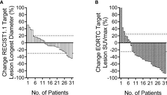
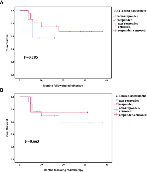
Results: Positron emission tomography-based local objective response rate was significantly higher than that based on computed tomography [70.97% (22/31) and 12.90% (4/31), respectively (p<0.001)]. The mean survival time of responders and non-responders assessed by positron emission tomography was 28.6 months vs. 11.4 months (p=0.29), whereas that assessed by computed tomography was 24.5 months vs. 26 months (p=0.66), respectively. Three radiomics features were screened to establish a personalized prediction nomogram with high area under curve (0.94, 95% CI 0.85-0.99, p<0.001). The decision curve showed a high clinical value of the radiomics nomogram.
Conclusions: We recommend positron emission tomography for evaluating the short-term efficacy of hypo-fractionated radiotherapy in non-small cell lung cancer, and that the radiomics nomogram could be an important technique for the prediction of short-term efficacy, which might enable an improved and precise treatment.
Registration number/url: ChiCTR1900027768/http://www.chictr.org.cn/showprojen.aspx?proj=46057.
Keywords: computed tomography; hypo-fractionated radiotherapy; non-small-cell lung cancer; positron emission tomography; radiomics.
Copyright © 2021 Jiang, Gao, Chen, Shi, Wu, Chen, Zhang, Pang and Lin.
Conflict of interest statement
The authors declare that the research was conducted in the absence of any commercial or financial relationships that could be construed as a potential conflict of interest.
Figures

Similar articles
-
A CT-based deep learning radiomics nomogram for predicting the response to neoadjuvant chemotherapy in patients with locally advanced gastric cancer: A multicenter cohort study.EClinicalMedicine. 2022 Mar 21;46:101348. doi: 10.1016/j.eclinm.2022.101348. eCollection 2022 Apr. EClinicalMedicine. 2022. PMID: 35340629 Free PMC article.
-
Development of a computed tomography-based radiomics nomogram for prediction of transarterial chemoembolization refractoriness in hepatocellular carcinoma.World J Gastroenterol. 2021 Jan 14;27(2):189-207. doi: 10.3748/wjg.v27.i2.189. World J Gastroenterol. 2021. PMID: 33510559 Free PMC article.
-
Development of a radiomics nomogram based on the 2D and 3D CT features to predict the survival of non-small cell lung cancer patients.Eur Radiol. 2019 May;29(5):2196-2206. doi: 10.1007/s00330-018-5770-y. Epub 2018 Dec 6. Eur Radiol. 2019. PMID: 30523451
-
The Promise and Future of Radiomics for Personalized Radiotherapy Dosing and Adaptation.Semin Radiat Oncol. 2023 Jul;33(3):252-261. doi: 10.1016/j.semradonc.2023.03.003. Semin Radiat Oncol. 2023. PMID: 37331780 Free PMC article. Review.
-
PET radiomics in lung cancer: advances and translational challenges.EJNMMI Phys. 2024 Oct 3;11(1):81. doi: 10.1186/s40658-024-00685-5. EJNMMI Phys. 2024. PMID: 39361110 Free PMC article. Review.
Cited by
-
A pilot trial of consolidation bevacizumab after hypo-fractionated concurrent chemoradiotherapy in patients with unresectable locally advanced non-squamous non-small-cell lung cancer.Cancer Med. 2023 Sep;12(17):17638-17647. doi: 10.1002/cam4.6381. Epub 2023 Aug 3. Cancer Med. 2023. PMID: 37537968 Free PMC article.
-
Noninvasive optoacoustic imaging of breast tumor microvasculature in response to radiotherapy.Front Physiol. 2022 Oct 17;13:1044308. doi: 10.3389/fphys.2022.1044308. eCollection 2022. Front Physiol. 2022. PMID: 36324309 Free PMC article.
-
Dynamic PET Imaging Using Dual Texture Features.Front Comput Neurosci. 2022 Jan 7;15:819840. doi: 10.3389/fncom.2021.819840. eCollection 2021. Front Comput Neurosci. 2022. PMID: 35069162 Free PMC article.
References
-
- Takeda A, Kunieda E, Fujii H, Yokosuka N, Aoki Y, Oooka Y, et al. . Evaluation for local failure by 18F-FDG PET/CT in comparison with CT findings after stereotactic body radiotherapy (SBRT) for localized non-small-cell lung cancer. Lung Cancer (2013) 79(3):248–53. 10.1016/j.lungcan.2012.11.008 - DOI - PubMed
LinkOut - more resources
Full Text Sources
Other Literature Sources