. 2021 Feb 26:11:625612.
doi: 10.3389/fonc.2021.625612. eCollection 2021.

Upregulation of PEDF Predicts a Poor Prognosis and Promotes Esophageal Squamous Cell Carcinoma Progression by Modulating the MAPK/ERK Signaling Pathway

Affiliations

Upregulation of PEDF Predicts a Poor Prognosis and Promotes Esophageal Squamous Cell Carcinoma Progression by Modulating the MAPK/ERK Signaling Pathway

Zui Chen et al. Front Oncol. .

Abstract

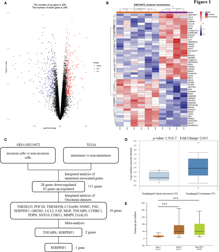
Invasion and metastasis represent the primary causes of therapeutic failure in patients diagnosed with esophageal squamous cell carcinoma (ESCC). The lack of effective treatment strategies for metastatic ESCC is the major cause of the low survival rate. Therefore, it is crucial to understand the molecular mechanisms underlying ESCC metastasis and identify potential biomarkers for targeted therapy. Herein, we reported that PEDF is significantly correlated with tumor cell invasion and metastasis in ESCC. The high expression of PEDF is an independent unfavorable prognostic factor for ESCC patients' overall survival (OS). We successfully developed and verified a nomogram to predict the preoperative OS of ESCC patients, and the actual and nomogram-predicted 1-, 3-, and 5-year survival rates had good consistency. The receiver operating characteristic (ROC) curve showed that the area under the curve (AUC) values for 1-, 3- and 5- survival were 0.764, 0.871, and 0.91, respectively. Overexpression of PEDF significantly promoted the migration and invasion of ESCC cells in vitro, while silencing PEDF yielded the opposite effects. Elevated levels of PEDF altered the expression of proteins involved in epithelial-mesenchymal transition (EMT), as indicated by the upregulation of N-cadherin and the downregulation of α-catenin and E-cadherin in ESCC cells. Mechanistically, PEDF promoted tumor cell motility and EMT by activating the MAPK/ERK signaling pathway. In conclusion, our results reveal that PEDF is involved in ESCC metastasis and could act as a prognostic factor for ESCC. Our research provides a fresh perspective into the mechanism of ESCC metastasis.

Keywords: MAPK/ERK; esophageal squamous cell carcinoma; metastasis; pigment epithelium-derived factor; prognosis.


Conflict of interest statement

The authors declare that the research was conducted in the absence of any commercial or financial relationships that could be construed as a potential conflict of interest.

Figures

Figure 1
Figure 1
PEDF expression is upregulated in esophageal carcinoma invasion cells. (A) A Pearson test was used to analyze genes differentially expressed in ESCC. (B) The cluster heat maps showing the 50 different expressed mRNAs in four lines of ESCC model invasion cells and corresponding six lines of ESCC model non-invasion cells. The blue and red stripes represent downregulated and upregulated mRNAs respectively. (C) Microarray analysis was performed to identify metastasis associated genes. Expression levels were analyzed in GEO (invasion cells vs non-invasion cells) and TCGA (metastasis vs non-metastasis). In order to rule out effects of precancerous lesions, esophageal cancer precursor genes and esophageal carcinoma genes were identified in Oncomine microarray dataset, as shown in the flowchart. These data were integrated with meta-analysis to identify the potential esophageal carcinoma metastasis associated genes (two genes). (D) Representative PEDF mRNA expression in esophageal carcinoma tissues versus esophageal cancer precursor samples from the Kim Esophagus dataset. (E) The significant association between clinic stages and PEDF expression levels. ***P < 0.001.
Figure 2
Figure 2
PEDF is abnormally expressed in advanced ESCC patients and related to clinical indicators. (A) Levels of PEDF protein expression in ESCC tissue chip are shown under both low and high magnifications. (B) The immunohistochemical score was calculated for each ESCC samples. Tumors with an expression level lower than six points are considered the low expression, while six points or more are considered high expression. (C) Graph showing the IHC staining scores and pathological clinical stage in 114 ESCC patients. **P < 0.01, ***P < 0.001. (D) Kaplan–Meier curve analysis revealed that ESCC patients in high PEDF expression had shorter survival time than that of ESCC patients with low expression of PEDF. The survival time was significantly associated with gender, T stage, N stage, TNM stage, but not with age, tumor grade, CD8, PD-L1.
Figure 3
Figure 3
A nomogram prediction model was constructed based on the expression of PEDF in ESCC. (A) Nomogram system for the 1-, 3-, and 5- year survival rate prediction. The nomogram prediction system was a novel model to estimate OS based on related factors (tumor grade, T stage, N stage, TNM), patient-specific factors (age, gender) and PEDF expression. (B) Calibration plots for predicting for ESCC OS at 1-, 3-, and 5-year. The blue dotted line indicates the ideal nomogram; blue X indicates the bootstrap-corrected estimates; vertical bars indicate the 95% CIs. (C) ROC curves of the 1-, 3-, and 5-year nomograms of ESCC patients. The red bars represent a new nomogram predicted OS, whereas the black bars represent the TNM stage predicted OS.
Figure 4
Figure 4
PEDF promotes ESCC cell migration, invasion, and EMT. (A) The KYSE510 and KYSE140 cellular migration and invasion capability were assessed by transwell migration and Boyden chamber invasion assays. The mean numbers of migrating or invading cells in the field were calculated by mean ± SD from three-time different experiments. Magnification for ×100. *P < 0.05, **P < 0.01, ***P < 0.001. (B) KYSE510 and KYSE140 cells and appropriate controls were used in a wound-healing assay. Scale bar = 100 μm. (n = 3 each). Data are means ± S.E.M. *P < 0.05, **P < 0.01. (C) The expression of α-catenin, N-cadherin, and E-cadherin were probed by western blotting in overexpress of PEDF KYSE510 and KYSE140 cells, and PEDF inhibition KYSE510 and KYSE140 cells, and the corresponding control cells.
Figure 5
Figure 5
MAPK/ERK signaling pathway is required for PEDF mediated EMT. (A) The expressions of AKT, p-AKT, a-catenin, ERK1/2, p-ERK1/2 and PEDF were probed by western blotting in the overexpression of PEDF KYSE510 and KYSE140 cells, and PEDF knockdown KYSE510 and KYSE140 cells, and the corresponding control cells. GAPDH was a protein loading control. (B) Western blotting indicated that p-ERK1/2 inhibitor PD98059 and U0126 inhibited PEDF induced the activation of MAPK/ERK signaling pathway, as well as the EMT-related characterizations in KYSE510 and KYSE140 cells. Cells were treated with MAPK/ERK signaling agonist PD98059 or U0126 for 24 h, and then performed WB assay. P < 0.05, relative to PD98059 or U0126 treatment of the same cell type as controls. (C) The cell invasion and migration were evaluated after ERK1/2 inhibitor treatment for 24 h. Magnification for × 100. *P < 0.05, **P < 0.01, ***P < 0.001.

Similar articles

Cited by

References

    1. Bray F, Ferlay J, Soerjomataram I, Siegel RL, Torre LA, Jemal A. Global cancer statistics 2018: GLOBOCAN estimates of incidence and mortality worldwide for 36 cancers in 185 countries. CA Cancer J Clin (2018) 68(6):394–424. 10.3322/caac.21492 - DOI - PubMed
    1. Chen W, Zheng R, Baade PD, Zhang S, Zeng H, Bray F, et al. . Cancer statistics in China. CA Cancer J Clin (2016) 66(2):115–32. 10.3322/caac.21338 - DOI - PubMed
    1. Enzinger PC, Mayer RJ. Esophageal Cancer. N Engl J Med (2003) 349: (23):2241–52. 10.1056/NEJMra035010 - DOI - PubMed
    1. Wijnhoven BP, Tran KT, Esterman A, Watson DI, Tilanus HW. An evaluation of prognostic factors and tumor staging of resected carcinoma of the esophagus. Ann Surg (2007) 245(5):717–25. 10.1097/01.sla.0000251703.35919.02 - DOI - PMC - PubMed
    1. Furihata T, Sakai T, Kawamata H, Omotehara F, Shinagawa Y, Imura J, et al. . A new in vivo model for studying invasion and metastasis of esophageal squamous cell carcinoma. Int J Oncol (2001) 19(5):903–7. 10.3892/ijo.19.5.903 - DOI - PubMed