. 2021 Feb 25:11:626748.
doi: 10.3389/fonc.2021.626748. eCollection 2021.

PD-L2 Is Constitutively Expressed in Normal and Malignant Urothelium

Affiliations

PD-L2 Is Constitutively Expressed in Normal and Malignant Urothelium

Alexander C Dowell et al. Front Oncol. .

Abstract

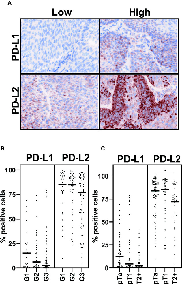
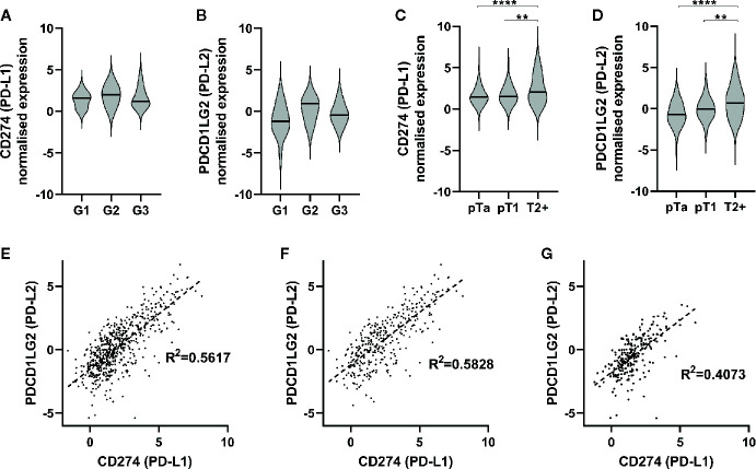
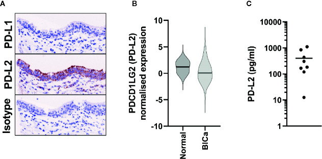
The use of immune checkpoint blockade, in particular PD-1 and PD-L1 inhibitors, is now commonplace in many clinical settings including the treatment of muscle-invasive bladder cancer (MIBC). Notwithstanding, little information exists regarding the expression of the alternative PD-1 ligand, PD-L2 in urothelial bladder cancer (UBC). We therefore set out to characterise the expression of PD-L2 in comparison to PD-L1. Firstly, we assessed PD-L2 expression by immunohistochemistry and found widespread expression of PD-L2 in UBC, albeit with reduced expression in MIBC. We further investigated these findings using RNA-seq data from a cohort of 575 patients demonstrating that PDCD1LG2 (PD-L2) is widely expressed in UBC and correlated with CD274 (PD-L1). However, in contrast to our immunohistochemistry findings, expression was significantly increased in advanced disease. We have also provided detailed evidence of constitutive PD-L2 expression in normal urothelium and propose a mechanism by which PD-L2 is cleaved from the cell surface in MIBC. These data provide a comprehensive assessment of PD-L2 in UBC, showing PD-L2 is abundant in UBC and, importantly, constitutively present in normal urothelium. These data have implications for future development of immune checkpoint blockade, and also the understanding of the function of the immune system in the normal urinary bladder.

Keywords: PD-L1 (B7-H1 CD274); PD-L2: programmed cell death ligand 2; bladder cancer; immune checkpoint inhibitors; normal urothelium.


Conflict of interest statement

RB has contributed to advisory boards for Olympus Medical Systems and Janssen, and undertakes research funded by UroGen Pharma, QED Therapeutics and Janssen. NJ has contributed to advisory boards for Merck USA and Pierre Fabre. The remaining authors declare that the research was conducted in the absence of any commercial or financial relationships that could be construed as a potential conflict of interest.

Figures

Figure 1
Figure 1
PD-L2 may be altered in muscle-invasive disease. PD-L1 and PD-L2 expression on tumor cells was investigated by immunohistochemistry staining of TMA. Staining was visualised by tyramine signal amplification. Data is presented from 123 and 146 patient samples, which were suitable for analysis, respectively. (A) Representative cases with high or low levels of PD-L1 and PD-L2 staining, as indicated. PD-1 ligand (as indicated) staining (brown), with nuclear counter stain (blue). (B, C) Positive immunohistochemical staining was assessed by inForm automated tissue analysis, and is presented as percentage positive tumor cells. Expression of PD-1 ligands is shown in respect of grade (B) and stage (C) One-way ANOVA with Tukey’s multiple comparison test; *p = 0.0296.
Figure 2
Figure 2
PD-1 ligands are expressed in urothelial bladder cancer (UBC), and are associated with increasing stage. (A, B) RNA-seq was performed on tumor samples from 44 non-muscle invasive bladder cancer patients (pTa-pT1) and normalised. CD274 (PD-L1) (A) and PDCD1LG2 (PD-L2) (B) gene expression is presented in respect of tumor grade (G1 n=15, G2 n=3, G3 n=26). (C, D) RNA-seq data from two publicly available data sets with tumor stage data were included for further analysis. Data shown for 575 patients (pTa n=170, pT1 n=66, T2+ n=339). CD274 (PD-L1) (C) and PDCD1LG2 (PD-L2) (D) gene expression is shown with respect to tumor stage. One-way ANOVA with Tukey’s multiple comparison test; **p = < 0.01, ****p = < 0.0001. (E–G) Correlation of CD274 and PDCD1LG2 gene expression from combined RNA-seq cohorts. (E) All UBC samples (n = 575), (G) MIBC (n = 339), (F) NMIBC, and (n = 236).
Figure 3
Figure 3
PD-L2 is constitutively expressed in Normal Bladder tissue. (A) Representative immunohistochemistry staining of either PD-L1 or PD-L2 on normal ureter urothelium from TMAs. (B) PDCD1LG2 (PD-L2) gene expression from TCGA data from normal bladder tissue (n = 19), in comparison to all bladder cancer samples (n = 575). (C) Urinary PD-L2 levels were determined by ELISA in mid-stream urine samples from 8 healthy volunteers.

Similar articles

Cited by

References

    1. Cancer Research UK . (2020) Bladder Cancer https://www.cancerresearchuk.org/health-professional/cancer-statistics/s... [Accessed May 15 2020]
    1. Ghatalia P, Zibelman M, Geynisman DM, Plimack E. Approved checkpoint inhibitors in bladder cancer: which drug should be used when? Ther Adv Med Oncol (2018) 10:1758835918788310. 10.1177/1758835918788310 - DOI - PMC - PubMed
    1. Stenehjem DD, Tran D, Nkrumah MA, Gupta S. PD1/PDL1 inhibitors for the treatment of advanced urothelial bladder cancer. OncoTarg Ther (2018) 11:5973–89. 10.2147/OTT.S135157 - DOI - PMC - PubMed
    1. Nurieva RI, Liu X, Dong C. Molecular mechanisms of T-cell tolerance. Immunol Rev (2011) 241:133–44. 10.1111/j.1600-065X.2011.01012.x - DOI - PMC - PubMed
    1. Derks S, Nason KS, Liao X, Stachler MD, Liu KX, Liu JB, et al. . Epithelial PD-L2 Expression Marks Barrett’s Esophagus and Esophageal Adenocarcinoma. Cancer Immunol Res (2015) 3:1123–9. 10.1158/2326-6066.CIR-15-0046 - DOI - PMC - PubMed