

Sophoridine Inhibits the Tumour Growth of Non-Small Lung Cancer by Inducing Macrophages M1 Polarisation via MAPK-Mediated Inflammatory Pathway
- PMID: 33718223
- PMCID: PMC7943889
- DOI: 10.3389/fonc.2021.634851
Sophoridine Inhibits the Tumour Growth of Non-Small Lung Cancer by Inducing Macrophages M1 Polarisation via MAPK-Mediated Inflammatory Pathway
Abstract
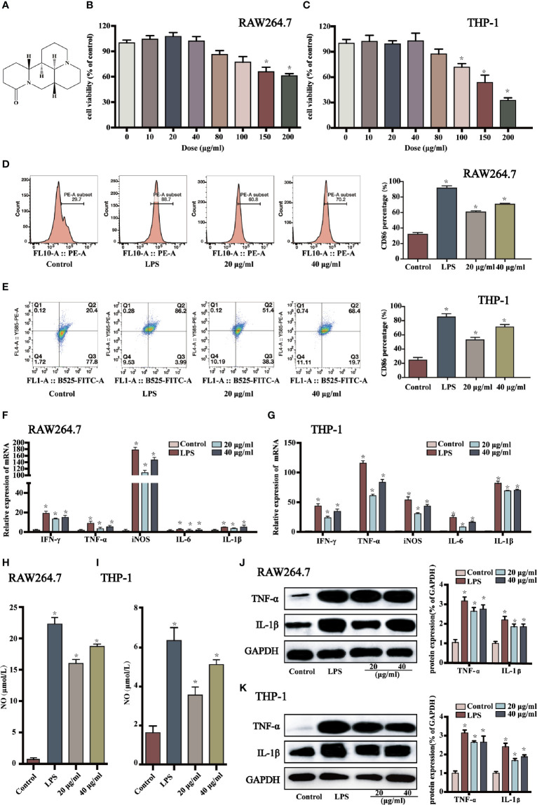
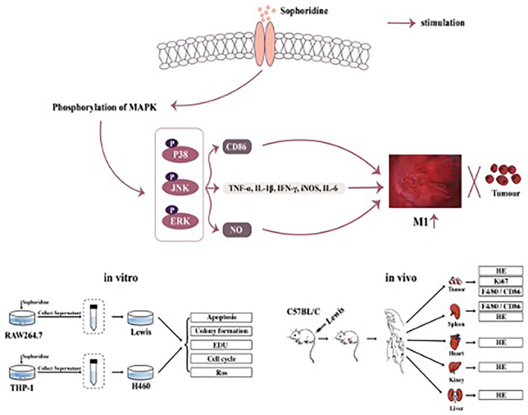
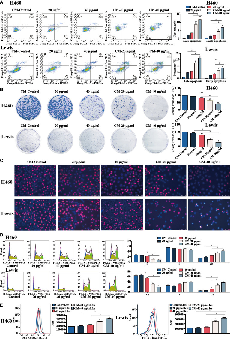
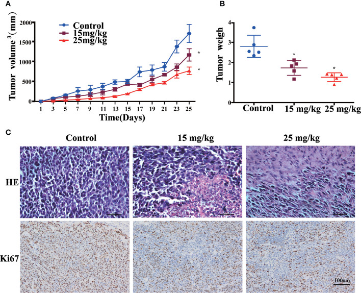
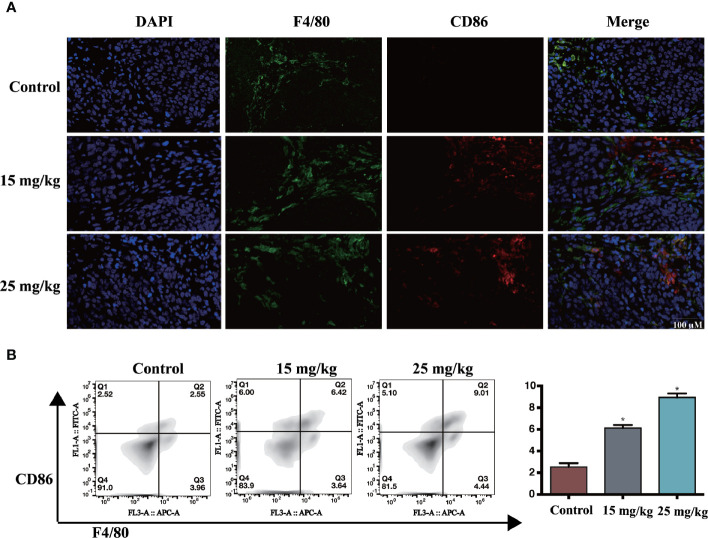
Lung cancer is one of the most common and lethal neoplasms for which very few efficacious treatments are currently available. M1-like polarised tumour-associated macrophages (TAMs) are key mediators to modulate the tumour microenvironment, which play a key role in inhibiting cancer cell growth. Sophoridine, a naturally occurring alkaloid, exerts multiple pharmacological activities including anti-tumour and anti-inflammatory activities, but it has not been characterised as a regulator of tumour microenvironment towards NSCLC. Herein, the regulatory effects of sophoridine on the polarisation of THP-1 cells into TAMs and the anti-tumour effects of sophoridine-stimulated M1 polarised macrophages towards lung cancer cells were carefully investigated both in vitro and in vivo. The results showed that sophoridine could significantly promote M1 polarisation of RAW264.7 and THP-1-derived macrophages, leading to increased expression of pro-inflammatory cytokines and the M1 surface markers CD86 via activating MAPKs signaling pathway. Further investigations showed that sophoridine-stimulated RAW264.7 and THP-1-derived M1 macrophages effectively induced cell apoptosis as well as inhibited the cell colony formation and cell proliferation in both H460 and Lewis lung cancer cells. In Lewis-bearing mice model, sophoridine (15 or 25 mg/kg) significantly inhibited the tumour growth and up-regulated the expression of CD86/F4/80 in tumour tissues. Collectively, the findings clearly demonstrate that sophoridine promoted M1-like polarisation in vitro and in vivo, suggesting that sophoridine held a great therapeutic potential for treating lung cancer.
Keywords: RAW264.7; THP-1 cells; macrophage differentiation; mitogen-activated protein kinase (MPKs) pathway; sophoridine.
Copyright © 2021 Zhao, Hui, Zeng, Yin, Huang, Tang, Ge and Lei.
Conflict of interest statement
The authors declare that the research was conducted in the absence of any commercial or financial relationships that could be construed as a potential conflict of interest.
Figures

Similar articles
-
Sophoridine suppresses macrophage-mediated immunosuppression through TLR4/IRF3 pathway and subsequently upregulates CD8+ T cytotoxic function against gastric cancer.Biomed Pharmacother. 2020 Jan;121:109636. doi: 10.1016/j.biopha.2019.109636. Epub 2019 Nov 13. Biomed Pharmacother. 2020. PMID: 31733580
-
Matrine suppresses lung cancer metastasis via targeting M2-like tumour-associated-macrophages polarization.Am J Cancer Res. 2021 Sep 15;11(9):4308-4328. eCollection 2021. Am J Cancer Res. 2021. PMID: 34659889 Free PMC article.
-
Modulation the crosstalk between tumor-associated macrophages and non-small cell lung cancer to inhibit tumor migration and invasion by ginsenoside Rh2.BMC Cancer. 2018 May 22;18(1):579. doi: 10.1186/s12885-018-4299-4. BMC Cancer. 2018. PMID: 29783929 Free PMC article.
-
The role of exosomes in the tumour microenvironment on macrophage polarisation.Biochim Biophys Acta Rev Cancer. 2022 Nov;1877(6):188811. doi: 10.1016/j.bbcan.2022.188811. Epub 2022 Oct 5. Biochim Biophys Acta Rev Cancer. 2022. PMID: 36208648 Review.
-
Tumour-associated macrophage polarisation and re-education with immunotherapy.Front Biosci (Elite Ed). 2015 Jan 1;7(2):293-308. doi: 10.2741/E735. Front Biosci (Elite Ed). 2015. PMID: 25553381 Review.
Cited by
-
Cordyceps sinensis relieves non-small cell lung cancer by inhibiting the MAPK pathway.Chin Med. 2024 Mar 25;19(1):54. doi: 10.1186/s13020-024-00895-0. Chin Med. 2024. PMID: 38528546 Free PMC article.
-
Sophoridine inhibits proliferation and migration by targeting PIM1 in breast cancer.Pharm Biol. 2025 Dec;63(1):503-523. doi: 10.1080/13880209.2025.2537123. Epub 2025 Jul 24. Pharm Biol. 2025. PMID: 40708207 Free PMC article.
-
Single-sample gene set enrichment analysis reveals the clinical implications of immune-related genes in ovarian cancer.Front Mol Biosci. 2024 Aug 5;11:1426274. doi: 10.3389/fmolb.2024.1426274. eCollection 2024. Front Mol Biosci. 2024. PMID: 39161779 Free PMC article.
-
Jiedu Xiaozheng Yin Inhibits the Progression of Colitis Associated Colorectal Cancer by Stimulating Macrophage Polarization Towards an M1 Phenotype via the TLR4 Pathway.Integr Cancer Ther. 2024 Jan-Dec;23:15347354241247061. doi: 10.1177/15347354241247061. Integr Cancer Ther. 2024. PMID: 38641964 Free PMC article.
-
Research progress of sophoridine's pharmacological activities and its molecular mechanism: an updated review.Front Pharmacol. 2023 Jun 16;14:1126636. doi: 10.3389/fphar.2023.1126636. eCollection 2023. Front Pharmacol. 2023. PMID: 37397472 Free PMC article. Review.
References
LinkOut - more resources
Full Text Sources
Other Literature Sources