Review
. 2021 Feb 24:11:608860.
doi: 10.3389/fcimb.2021.608860. eCollection 2021.

Interesting Biochemistries in the Structure and Function of Bacterial Effectors

Affiliations
Review

Interesting Biochemistries in the Structure and Function of Bacterial Effectors

Hazel Mak et al. Front Cell Infect Microbiol. .

Abstract

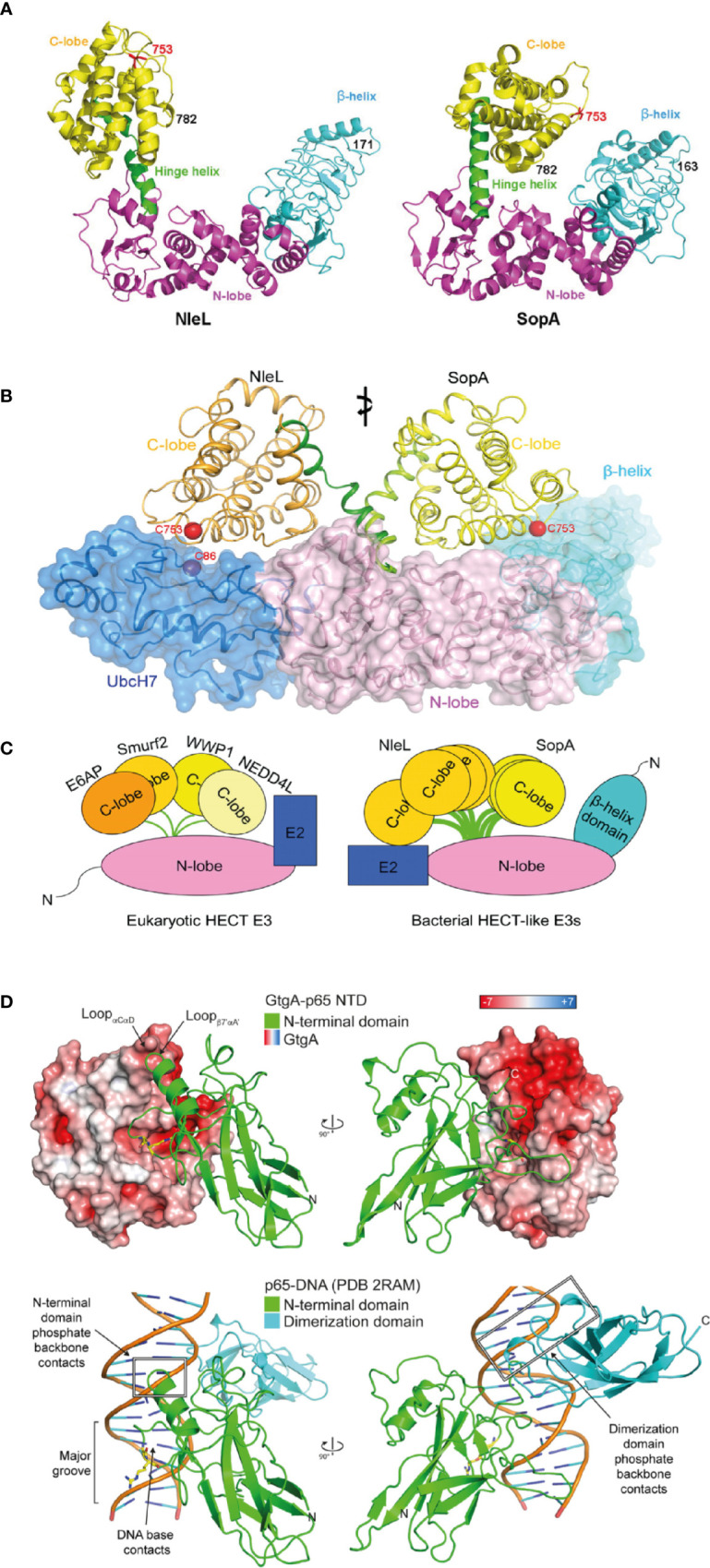
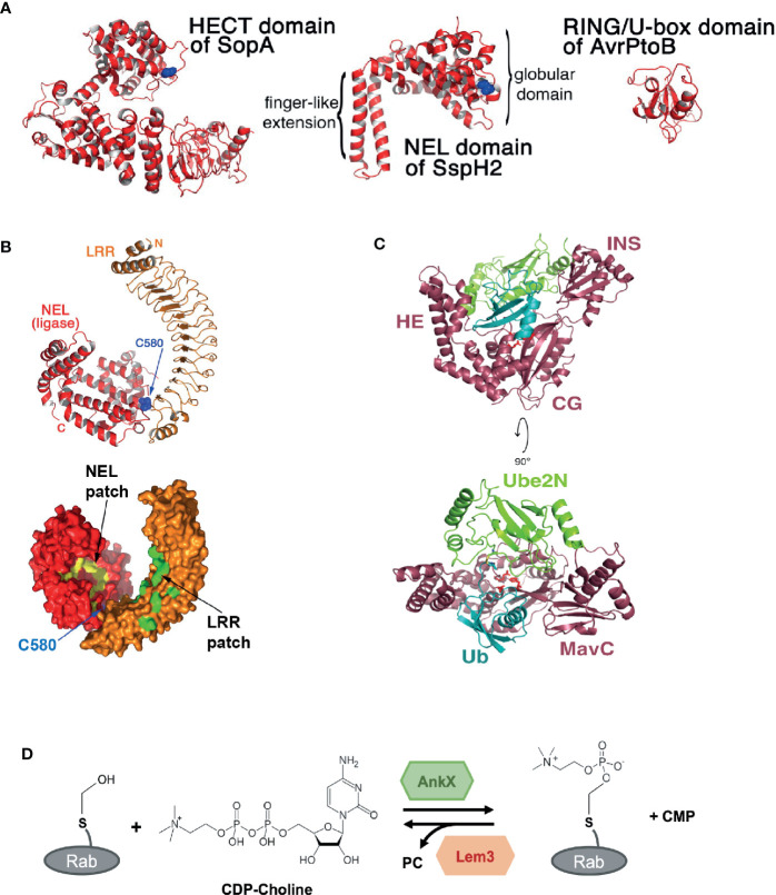
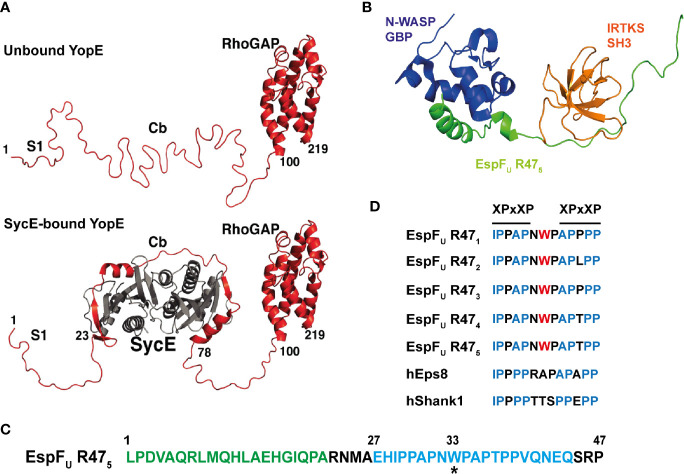
Bacterial effector proteins, delivered into host cells by specialized multiprotein secretion systems, are a key mediator of bacterial pathogenesis. Following delivery, they modulate a range of host cellular processes and functions. Strong selective pressures have resulted in bacterial effectors evolving unique structures that can mimic host protein biochemical activity or enable novel and distinct biochemistries. Despite the protein structure-function paradigm, effectors from different bacterial species that share biochemical activities, such as the conjugation of ubiquitin to a substrate, do not necessarily share structural or sequence homology to each other or the eukaryotic proteins that carry out the same function. Furthermore, some bacterial effectors have evolved structural variations to known protein folds which enable different or additional biochemical and physiological functions. Despite the overall low occurrence of intrinsically disordered proteins or regions in prokaryotic proteomes compared to eukaryotes proteomes, bacterial effectors appear to have adopted intrinsically disordered regions that mimic the disordered regions of eukaryotic signaling proteins. In this review, we explore examples of the diverse biochemical properties found in bacterial effectors that enable effector-mediated interference of eukaryotic signaling pathways and ultimately support pathogenesis. Despite challenges in the structural and functional characterisation of effectors, recent progress has been made in understanding the often unusual and fascinating ways in which these virulence factors promote pathogenesis. Nevertheless, continued work is essential to reveal the array of remarkable activities displayed by effectors.

Keywords: bacterial effectors; host-pathogen; pathogenesis; protein organization; secretion systems; structure-function.


Conflict of interest statement

The authors declare that the research was conducted in the absence of any commercial or financial relationships that could be construed as a potential conflict of interest.

Figures

Figure 1
Figure 1
Structured bacterial effectors mimicking host cell proteins. (A) Structural comparison between Enterohemorrhagic E. coli (EHEC) effector NleL (residue 170-782) and Salmonella effector SopA (residue 163-782) in ribbon cartoon representation. Structures consist of the N-lobe (magenta), the C-lobe (yellow) and the β-helix domain (cyan). The catalytic cysteine (Cys) residue 753 are labelled in red. The hinge helix is labelled and shown in green (taken from Lin et al., 2011). (B) Structural superposition of two bacterial HECT-like E3 ligases, SopA from Salmonella and NleL from EHEC, bound to human E2 protein UbcH7 (shown in dark blue). The E3 ligase N-lobes and β-helix domains are shown in pink and light blue respectively, as ribbon representation and transparent surface. The C-lobe is structurally flexible and shown in orange for NleL and in yellow for SopA. The catalytic cysteine (Cys) residues are shown in red (taken from Lin et al., 2012). (C) Schematic of structural comparison between eukaryotic HECT E3 ligases and bacterial HECT-like E3 ligases. Eukaryotic HECT E3 ligases include E6AP, Smurf2, WWP1, and NEDD4L. Bacterial HECT-like E3 ligases include SopA from Salmonella and NleL from EHEC. Structural flexibility is shown in the C-lobe of NleL and SopA (taken from Lin et al., 2012). (D) Structural mimicry of DNA by Salmonella effector GtgA. Top structure shows GtgA in complex with the N-terminal domain (NTD) of p65. Bottom structure shows DNA in complex with the NTD and the dimerization domain of p65. GtgA is shown in surface representation and coloured according to its electrostatic surface potential (red is negative; white is neutral; blue is positive). The NTD of p65 is shown in green and the dimerization domain of p65 is shown in cyan. The cleavage site residues in p65 (Gly-40/Arg-41) are shown with yellow sticks (taken from Jennings et al., 2018).
Figure 2
Figure 2
Novel structures and biochemical activities mediated by bacterial effectors. (A) Structural comparison between the Novel E3 Ligase (NEL) domain in Salmonella effector SspH2 with bacterial E3 ligases that mimic eukaryotic E3 ligase domains: the homologous to E6-AP carboxy terminus (HECT) domain in SopA from Salmonella and the Really Interesting New Gene (RING/U-box) domain in AvrPtoB from Pseudomonas Syringae. Catalytic cysteine residues are shown in blue (taken from Quezada et al., 2009). (B) Crystal structure of Salmonella effector SspH2 shown in ribbon representation (top) and molecular surface representation (bottom). The Novel E3 Ligase (NEL) domain is shown in red and the leucine-rich repeat (LRR) domain in orange. The catalytic cysteine residue in SspH2 (C580) is shown in blue. Hydrophobic patches are labelled and shown in yellow for the NEL domain and in green for the LRR domain (taken and adapted from Quezada et al., 2009). (C) Crystal structure of the Legionella effector MavC bound to E2 Ube2N-ubiquitin conjugate. MavC is shown in dark pink, Ube2N in green and ubiquitin (Ub) in blue. The domains of MavC are labelled: helical extension (HE), core globular domain (CG), and insertion domain (INS). The active site residues in MavC (C74, H231, and Q252) are shown as red sticks (taken from Puvar et al., 2020). (D) Phosphocholination and dephosphocholination of Rab GTPase protein by the Legionella effector AnkX and Lem3. AnkX catalyzes phosphocholination, the transfer of the phosphocholine moiety from cytidine diphosphate (CDP)-choline onto the hydroxyl group of a serine residue in certain Rab GTPase proteins. Lem3 catalyzes the dephosphocholination by removing the phosphocholine (PC) (adapted from Heller et al., 2015).
Figure 3
Figure 3
Intrinsic disorder in bacterial effector proteins. (A) Crystal structure of Yersinia YopE (red) as unbound ‘free’ and bound to the chaperone SycE (grey) in ribbon representation. Functional regions of YopE are labelled: N-terminal secretion signal 1 (S1), chaperone-binding (Cb) and Rho-GAP domain. Intrinsic disorder occurs in the first 100 residues of YopE, which includes the S1 and Cb regions. The chaperone, SycE (gray), binds to the Cb region and cause disorder-to-order conformational change in the Cb region of YopE (taken and adapted from Rodgers et al., 2008). (B) Structure of the tri-molecular complex consisting of the GTPase binding domain (GBD) domain of N-WASP, the fifth consecutive 47-residue repeat of EspFU (EspFU R475) and the SH3 domain of IRTKS at the lowest energy conformation in ribbon representation. Structure obtained from NMR spectroscopy. N-WASP GBD, EspFU R475, and IRTKS SH3 are shown and labelled in dark blue, green and orange, respectively (PDB accession number 2LNH from Aitio et al., 2012). (C) Amino acid sequence of the fifth repeat of EspFU (EspFU R475). This repeat is one of the highly conserved consecutive 47-residue repeats in EspFU. The GBD domain of N-WASP interacts and binds to the N-terminal helix binding region shown in green, and the SH3 domain of IRTKS binds to the C-terminal Proline-rich region shown in blue. The asterisk (*) indicates the tryptophan switch in the linker region (adapted from Aitio et al., 2012). (D) Sequence alignment of the linker between the two XPxXP motif in the EspFU repeats and in other IRTKS binding interaction partners, including human Eps8 (hEps8) and human Shank1, showing the tryptophan switch in the linker region. In the XPxXP motif, “X” is a hydrophobic residue and “x” is any residue and P is proline (adapted from Aitio et al., 2012).

Similar articles

Cited by

References

    1. Aepfelbacher M., Roppenser B., Hentschke M., Ruckdeschel K. (2011). Activity Modulation of the Bacterial Rho GAP YopE: An Inspiration for the Investigation of Mammalian Rho GAPs. Eur. J. Cell Biol. 90 (11), 951–954. 10.1016/j.ejcb.2010.12.004 - DOI - PubMed
    1. Aitio O., Hellman M., Skehan B., Kesti T., Leong J. M., Saksela K., et al. . (2012). Enterohaemorrhagic Escherichia Coli Exploits a Tryptophan Switch to Hijack Host F-Actin Assembly. Structure 20 (10), 1692–1703. 10.1016/j.str.2012.07.015 - DOI - PMC - PubMed
    1. Akturk A., Wasilko D. J., Wu X., Liu Y., Zhang Y., Qiu J., et al. . (2018). Mechanism of Phosphoribosyl-Ubiquitination Mediated by a Single Legionella Effector. Nature 55 (7707), 729–745. 10.1038/s41586-018-0147-6 - DOI - PMC - PubMed
    1. Alto N. M., Shao F., Lazar C. S., Brost R. L., Chua G., Mattoo S., et al. . (2006). Identification of a Bacterial Type III Effector Family with G Protein Mimicry Functions. Cell 124 (1), 133–145. 10.1016/j.cell.2005.10.031 - DOI - PubMed
    1. Arbeloa A., Bulgin R. R., Mackenzie G., Shaw R. K., Pallen M. J., Crepin V. F., et al. . (2008). Subversion of Actin Dynamics by EspM Effectors of Attaching and Effacing Bacterial Pathogens. Cell. Microbiol. 10 (7), 1429–1441. 10.1111/j.1462-5822.2008.01136.x - DOI - PMC - PubMed

Publication types

LinkOut - more resources