

Higher-Order Chromatin Structures of Chromosomally Integrated HHV-6A Predict Integration Sites
- PMID: 33718266
- PMCID: PMC7953476
- DOI: 10.3389/fcimb.2021.612656
Higher-Order Chromatin Structures of Chromosomally Integrated HHV-6A Predict Integration Sites
Abstract
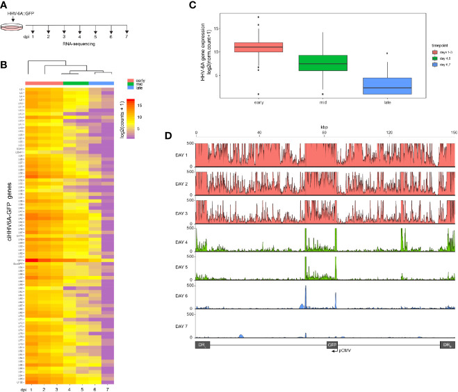
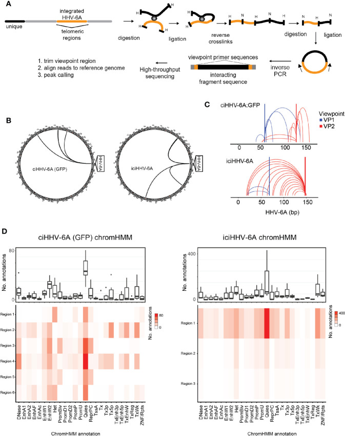
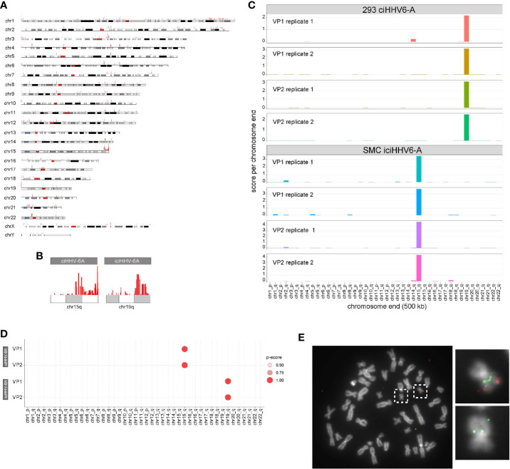
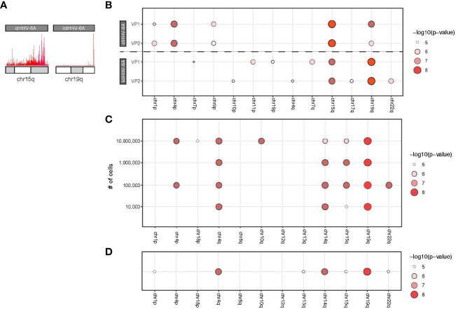
Human herpesvirus -6A and 6B (HHV-6A/B) can integrate their genomes into the telomeres of human chromosomes. Viral integration can occur in several cell types, including germinal cells, resulting in individuals that harbor the viral genome in every cell of their body. The integrated genome is efficiently silenced but can sporadically reactivate resulting in various clinical symptoms. To date, the integration mechanism and the subsequent silencing of HHV-6A/B genes remains poorly understood. Here we investigate the genome-wide chromatin contacts of the integrated HHV-6A in latently-infected cells. We show that HHV-6A becomes transcriptionally silent upon infection of these cells over the course of seven days. In addition, we established an HHV-6-specific 4C-seq approach, revealing that the HHV-6A 3D interactome is associated with quiescent chromatin states in cells harboring integrated virus. Furthermore, we observed that the majority of virus chromatin interactions occur toward the distal ends of specific human chromosomes. Exploiting this finding, we established a 4C-seq method that accurately detects the chromosomal integration sites. We further implement long-read minION sequencing in the 4C-seq assay and developed a method to identify HHV-6A/B integration sites in clinical samples.
Keywords: chromatin 3D architecture; epigenetics; gene expression; herpesvirus (hhv-6); latency.
Copyright © 2021 Mariani, Zimmerman, Rodriguez, Hasenohr, Aimola, Gerrard, Richman, Dest, Flamand, Kaufer and Frietze.
Conflict of interest statement
The authors declare that the research was conducted in the absence of any commercial or financial relationships that could be construed as a potential conflict of interest.
Figures
Similar articles
-
Inherited Chromosomally Integrated Human Herpesvirus 6 Demonstrates Tissue-Specific RNA Expression In Vivo That Correlates with an Increased Antibody Immune Response.J Virol. 2019 Dec 12;94(1):e01418-19. doi: 10.1128/JVI.01418-19. Print 2019 Dec 12. J Virol. 2019. PMID: 31597766 Free PMC article.
-
Inherited Chromosomally Integrated Human Herpesvirus 6 Genomes Are Ancient, Intact, and Potentially Able To Reactivate from Telomeres.J Virol. 2017 Oct 27;91(22):e01137-17. doi: 10.1128/JVI.01137-17. Print 2017 Nov 15. J Virol. 2017. PMID: 28835501 Free PMC article. Clinical Trial.
-
HHV-6A/B Integration and the Pathogenesis Associated with the Reactivation of Chromosomally Integrated HHV-6A/B.Viruses. 2017 Jun 26;9(7):160. doi: 10.3390/v9070160. Viruses. 2017. PMID: 28672870 Free PMC article. Review.
-
The Telomeric Repeats of Human Herpesvirus 6A (HHV-6A) Are Required for Efficient Virus Integration.PLoS Pathog. 2016 May 31;12(5):e1005666. doi: 10.1371/journal.ppat.1005666. eCollection 2016 May. PLoS Pathog. 2016. PMID: 27244446 Free PMC article.
-
Chromosomal Integration by Human Herpesviruses 6A and 6B.Adv Exp Med Biol. 2018;1045:209-226. doi: 10.1007/978-981-10-7230-7_10. Adv Exp Med Biol. 2018. PMID: 29896669 Review.
Cited by
-
Circular Chromosome Conformation Capture (4C-Seq) Analysis of HBV-Host Chromosome DNA Interactome.Methods Mol Biol. 2024;2837:45-58. doi: 10.1007/978-1-0716-4027-2_5. Methods Mol Biol. 2024. PMID: 39044074
-
Genomic and Epidemiological Investigations Reveal Chromosomal Integration of the Acipenserid Herpesvirus 3 Genome in Lake Sturgeon Acipenser fulvescens.Viruses. 2025 Apr 5;17(4):534. doi: 10.3390/v17040534. Viruses. 2025. PMID: 40284977 Free PMC article.
-
Long-read sequencing to detect full-length protein-protein interactions.Sci Rep. 2025 Jul 17;15(1):25967. doi: 10.1038/s41598-025-08549-3. Sci Rep. 2025. PMID: 40676061 Free PMC article.
-
Excision of Integrated Human Herpesvirus 6A Genomes Using CRISPR/Cas9 Technology.Microbiol Spectr. 2023 Mar 16;11(2):e0076423. doi: 10.1128/spectrum.00764-23. Online ahead of print. Microbiol Spectr. 2023. PMID: 36926973 Free PMC article.
-
Evasion of the Host Immune Response by Betaherpesviruses.Int J Mol Sci. 2021 Jul 13;22(14):7503. doi: 10.3390/ijms22147503. Int J Mol Sci. 2021. PMID: 34299120 Free PMC article. Review.
References
-
- Arbuckle J. H., Medveczky M. M., Luka J., Hadley S. H., Luegmayr A., Ablashi D., et al. . (2010). The latent human herpesvirus-6A genome specifically integrates in telomeres of human chromosomes in vivo and in vitro. Proc. Natl. Acad. Sci. U. S. A. 107 (12), 5563–5568. 10.1073/pnas.0913586107 - DOI - PMC - PubMed
-
- Boyd J. (2020). seqsetvis: Set Based Visualizations for Next-Gen Sequencing Data. R package version 1.8.0. ed.). Available at: https://bioconductor.org/packages/release/bioc/html/seqsetvis.html.
Publication types
MeSH terms
Substances
Grants and funding
LinkOut - more resources
Full Text Sources
Other Literature Sources
Molecular Biology Databases
Research Materials