. 2021 Feb 25:9:609311.
doi: 10.3389/fcell.2021.609311. eCollection 2021.

Clathrin Heavy Chain 1 Plays Essential Roles During Oocyte Meiotic Spindle Formation and Early Embryonic Development in Sheep

Affiliations

Clathrin Heavy Chain 1 Plays Essential Roles During Oocyte Meiotic Spindle Formation and Early Embryonic Development in Sheep

Zhe Han et al. Front Cell Dev Biol. .

Abstract

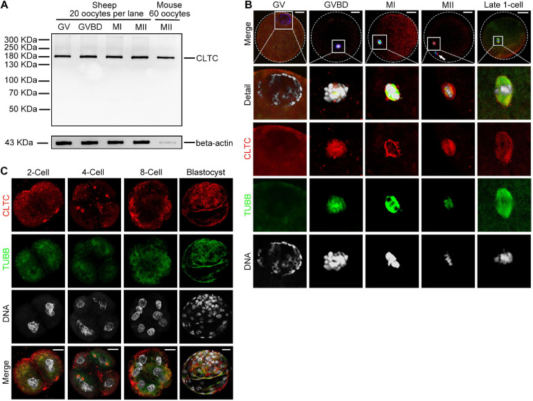
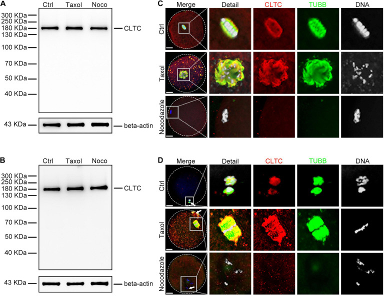
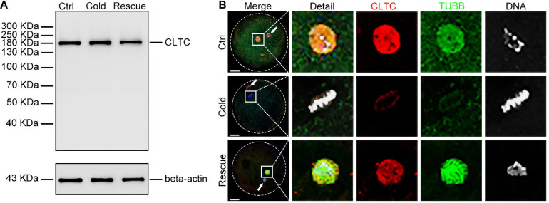
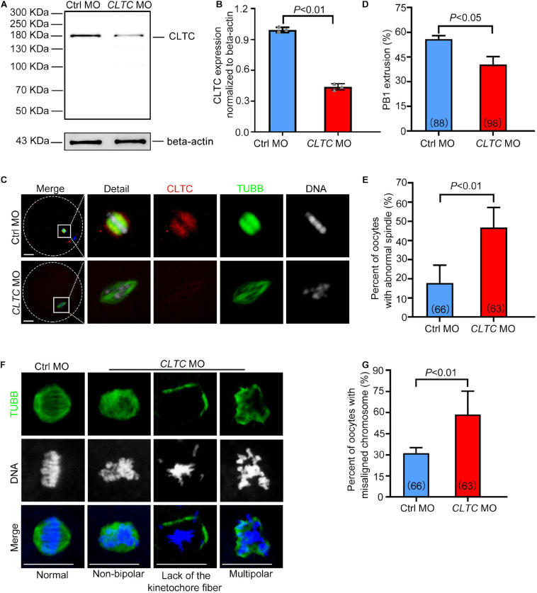
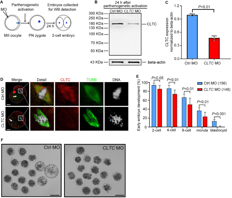
As a major protein of the polyhedral coat of coated pits and vesicles, clathrin molecules have been shown to play a stabilization role for kinetochore fibers of the mitotic spindle by acting as inter-microtubule bridges. Clathrin heavy chain 1 (CLTC), the basic subunit of the clathrin coat, plays vital roles in both spindle assembly and chromosome congression during somatic-cell mitosis. However, its function in oocyte meiotic maturation and early embryo development in mammals, especially in domesticated animals, has not been fully investigated. In this study, the expression profiles and functional roles of CLTC in sheep oocytes were investigated. Our results showed that the expression of CLTC was maintained at a high level from the germinal vesicle (GV) stage to metaphase II stage and that CLTC was distributed diffusely in the cytoplasm of cells at interphase, from the GV stage to the blastocyst stage. After GV breakdown (GVBD), CLTC co-localized with beta-tubulin during metaphase. Oocyte treatments with taxol, nocodazole, or cold did not affect CLTC expression levels but led to disorders of its distribution. Functional impairment of CLTC by specific morpholino injections in GV-stage oocytes led to disruptions in spindle assembly and chromosomal alignment, accompanied by impaired first polar body (PB1) emissions. In addition, knockdown of CLTC before parthenogenetic activation disrupted spindle formation and impaired early embryo development. Taken together, the results demonstrate that CLTC plays a vital role in sheep oocyte maturation via the regulation of spindle dynamics and an essential role during early embryo development.

Keywords: CLTC; chromosome congression; early embryo development; oocyte; spindle assembly.


Conflict of interest statement

The authors declare that the research was conducted in the absence of any commercial or financial relationships that could be construed as a potential conflict of interest.

Figures

FIGURE 1
FIGURE 1
Expression and subcellular localization of CLTC during sheep oocyte meiotic maturation and early embryonic development. (A) Immunoblotting analysis of CLTC in sheep oocytes during different stages of maturation (GV, GVBD, MI, and MII) and in mouse oocytes at MII stage. Lysate from 20 sheep oocytes or 60 mouse oocytes was loaded in the corresponding lanes. Beta-actin was used as a loading control. Oocyte samples were collected after being cultured for 0, 8, 12, and 24 h to reach the stages of GV, GVBD, MI, and MII, respectively. (B,C) Subcellular localizations of CLTC at each stage of oocyte and early embryonic development were detected by immunofluorescence staining. Slides were examined using a confocal microscope. White arrow indicates a polar body. Red, CLTC; green, TUBB; blue, DNA. Scale bar = 20 μm. CLTC, clathrin heavy chain 1; GV, germinal vesicle; GVBD, germinal vesicle breakdown; MI, metaphase I; MII, metaphase II.
FIGURE 2
FIGURE 2
Expression and localization of CLTC in metaphase oocytes treated with either taxol or nocodazole. (A) Immunoblotting analysis of CLTC from MI-stage oocytes after treatment with taxol or nocodazole. Beta-actin was used as a loading control. Noco, nocodazole. (B) Immunoblotting analysis of CLTC from MII-stage oocytes after treatment with either taxol or nocodazole. Beta-actin was used as a loading control. Noco, nocodazole. (C) Subcellular localizations of CLTC in MI-stage oocytes after treatment with either taxol or nocodazole were detected by immunofluorescence staining, respectively. Slides were examined using a confocal microscope. Red, CLTC; green, TUBB; blue, DNA. Scale bar = 20 μm. (D) Subcellular localizations of CLTC in MII-stage oocytes after treatment with either taxol or nocodazole were detected by immunofluorescence staining, respectively. Slides were examined using a confocal microscope. White arrows indicate polar bodies. Red, CLTC; green, TUBB; blue, DNA. Scale bar = 20 μm. CLTC, clathrin heavy chain 1; MI, metaphase I; MII, metaphase II.
FIGURE 3
FIGURE 3
Expression and localization of CLTC in MII oocytes after cold treatment. (A) Immunoblotting analysis of CLTC from MII-stage oocytes after cold or rescue treatment. Beta-actin was used as a loading control. (B) Subcellular localizations of CLTC in MII-stage oocytes after cold or rescue treatment were detected by immunofluorescence staining. Slides were examined using a confocal microscope. White arrows indicate polar bodies. Red, CLTC; green, TUBB; blue, DNA. Scale bar = 20 μm. CLTC, clathrin heavy chain 1; MII, metaphase II.
FIGURE 4
FIGURE 4
CLTC knockdown in sheep oocytes induces spindle defects and misaligned chromosomes. (A) Immunoblotting of CLTC expression after either CLTC-MO or control-MO injections. Beta-actin was used as a loading control. (B) Band intensities (representing CLTC expression) were assessed using gray-level analyses after CLTC-MO or control-MO injections. (C) The CLTC, spindles, and DNA in MII-stage oocytes (control-MO and CLTC-MO groups) were stained with anti-CLTC (red), anti-tubulin (green), and DAPI (blue), respectively. Scale bar = 20 μm. (D) The percentage of oocytes demonstrating PB1 extrusion in the control-MO and CLTC-MO groups. (E) Percentages of oocytes with abnormal spindles in the control-MO and CLTC-MO groups. (F) Immunofluorescence staining of CLTC expression after CLTC-MO or control-MO injections. Green, TUBB; blue, DNA. Scale bar = 20 μm. (G) Percentages of oocytes with chromosome misalignments in the control-MO and CLTC-MO groups. Data are presented as mean ± SD from at least three independent experiments. P < 0.05 was considered statistically significant. CLTC, clathrin heavy chain 1; MO, morpholino; PB1, first polar body; DAPI, 4′,6-diamidino-2-phenylindole.
FIGURE 5
FIGURE 5
MII-stage knockdown of CLTC impairs embryonic development. (A) Scheme for sample collections for immunoblotting analyses after MO injections into MII-stage oocytes. (B) CLTC-expression immunoblotting after CLTC-MO or control-MO injections into MII-stage sheep oocytes. Beta-actin was used as a loading control. (C) Band intensities (representing CLTC expression) were assessed using gray-level analyses after CLTC-MO or control-MO injections in MII-stage sheep oocytes. Data are presented as mean ± SD from three independent experiments. P < 0.05 was considered statistically significant. (D) The CLTC, spindles, and DNA in two-cell embryos from the control-MO and CLTC-MO groups were stained with anti-CLTC (red), anti-tubulin (green), and DAPI (blue), respectively. Scale bar = 20 μm. White arrows indicate the other blastomere nucleus. (E) MII-stage sheep oocytes were injected with either CLTC-MO or control-MO, and then the rate of early embryo developmental was measured. Data are presented as mean ± SD from eight independent repeats, and at least 12 MII oocytes were included for parthenogenetic activation in each group for each repeat. P < 0.05 was considered statistically significant. (F) Representative images of early embryos after CLTC-MO or control-MO injections. Scale bar = 100 μm. MII, metaphase II; CLTC, clathrin heavy chain 1; MO, morpholino; DAPI, 4′,6-diamidino-2-phenylindole.

Similar articles

References

    1. Booth D. G., Hood F. E., Prior I. A., Royle S. J. (2011). A TACC3/ch-TOG/clathrin complex stabilises kinetochore fibres by inter-microtubule bridging. EMBO J. 30 906–919. 10.1038/emboj.2011.15 - DOI - PMC - PubMed
    1. Cao Y. N., Zheng L. L., Wang D., Liang X. X., Gao F., Zhou X. L. (2018). Recent advances in microtubule-stabilizing agents. Eur. J. Med. Chem. 143 806–828. 10.1016/j.ejmech.2017.11.062 - DOI - PubMed
    1. Cheeseman L. P., Booth D. G., Hood F. E., Prior I. A., Royle S. J. (2011). Aurora a kinase activity is required for localization of TACC3/ch-TOG/clathrin inter-microtubule bridges. Commun. Integr. Biol. 4 409–412. 10.4161/cib.15250 - DOI - PMC - PubMed
    1. Cheeseman L. P., Harry E. F., Mcainsh A. D., Prior I. A., Royle S. J. (2013). Specific removal of TACC3-ch-TOG-clathrin at metaphase deregulates kinetochore fiber tension. J. Cell Sci. 126 2102–2113. 10.1242/jcs.124834 - DOI - PMC - PubMed
    1. Cotterill M., Harris S. E., Collado Fernandez E., Lu J., Huntriss J. D., Campbell B. K., et al. (2013). The activity and copy number of mitochondrial DNA in ovine oocytes throughout oogenesis in vivo and during oocyte maturation in vitro. Mol. Hum. Reprod. 19 444–450. 10.1093/molehr/gat013 - DOI - PMC - PubMed