. 2021 Feb 25:9:625540.
doi: 10.3389/fcell.2021.625540. eCollection 2021.

SIRT6 Maintains Redox Homeostasis to Promote Porcine Oocyte Maturation

Affiliations

SIRT6 Maintains Redox Homeostasis to Promote Porcine Oocyte Maturation

Yu Li et al. Front Cell Dev Biol. .

Abstract

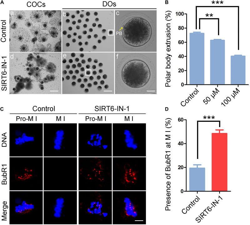
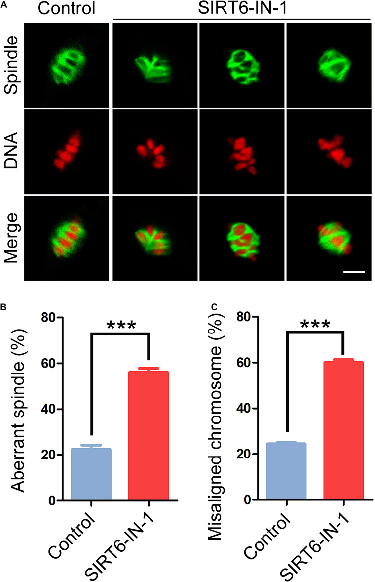
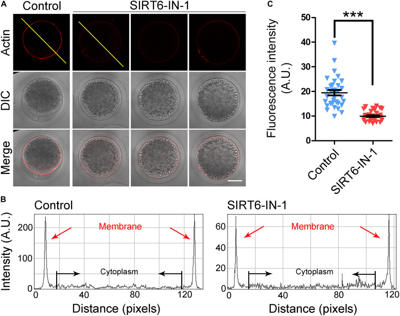
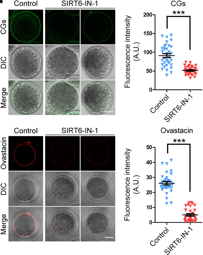
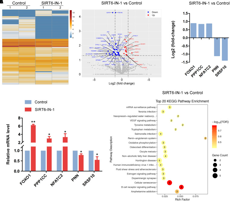
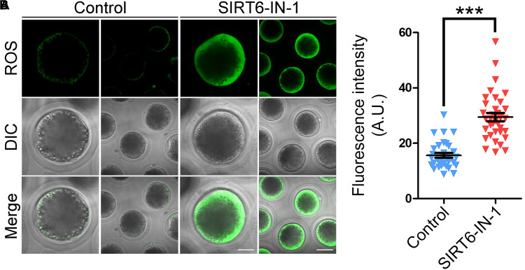
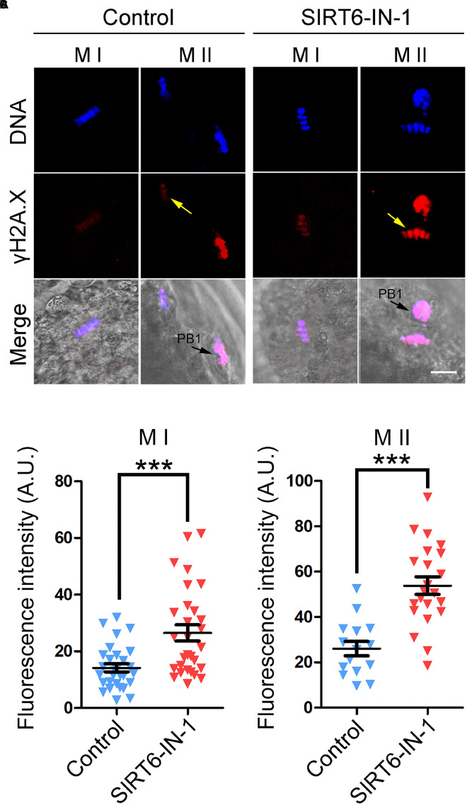
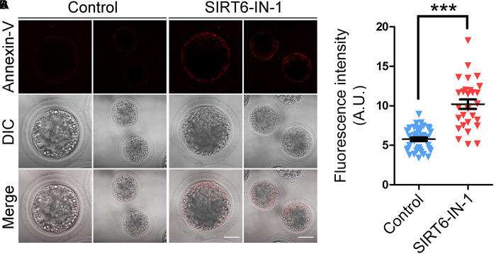
SIRT6, the sixth member of the sirtuin family proteins, has been characterized as a crucial regulator in multiple molecular pathways related to aging, including genome stability, DNA damage repair, telomere maintenance, and inflammation. However, the exact roles of SIRT6 during female germ cell development have not yet been fully determined. Here, we assessed the acquisition of meiotic competency of porcine oocytes by inhibition of SIRT6 activity. We observed that SIRT6 inhibition led to the oocyte meiotic defects by showing the impairment of polar body extrusion and cumulus cell expansion. Meanwhile, the compromised spindle/chromosome structure and actin dynamics were also present in SIRT6-inhibited oocytes. Moreover, SIRT6 inhibition resulted in the defective cytoplasmic maturation by displaying the disturbed distribution dynamics of cortical granules and their content ovastacin. Notably, we identified that transcript levels of genes related to oocyte meiosis, oxidative phosphorylation, and cellular senescence were remarkably altered in SIRT6-inhibited oocytes by transcriptome analysis and validated that the meiotic defects caused by SIRT6 inhibition might result from the excessive reactive oxygen species (ROS)-induced early apoptosis in oocytes. Taken together, our findings demonstrate that SIRT6 promotes the porcine oocyte meiotic maturation through maintaining the redox homeostasis.

Keywords: SIRT6; apoptosis; meiotic failure; oocyte maturation; redox homeostasis.


Conflict of interest statement

The authors declare that the research was conducted in the absence of any commercial or financial relationships that could be construed as a potential conflict of interest.

Figures

FIGURE 1
FIGURE 1
Effect of SIRT6 inhibition on meiotic maturation and spindle assembly checkpoint (SAC) activation in porcine oocytes. (A) Representative images of in vitro-matured oocytes in control and SIRT6-inhibited groups. Polar body extrusion (PBE) of denuded oocytes (DOs) and cumulus cell expansion of cumulus cell–oocyte complexes (COCs) were imaged by the confocal microscope. Scale bar, 360 μm (a,d); 240 μm (b,e); 30 μm (c,f). (B) The proportion of PBE was calculated in control and different concentrations of SIRT6-inhibited groups (50 and 100 μM) after 44 h in vitro culture. (C) The localization of BubR1 on the chromosomes in control and SIRT6-inhibited oocytes at pro-metaphase I (M I) and M I stages. Porcine oocytes were immunostained with BubR1 antibody and counterstained with Hoechst. Scale bar, 10 μm. (D) The proportion of BubR1 presence at the M I stage was calculated in control and SIRT6-inhibited oocytes. Data of panels (B,D) were shown as the mean percentage (mean ± SEM) of at least three independent experiments. **P < 0.01, ***P < 0.001.
FIGURE 2
FIGURE 2
Effect of SIRT6 inhibition on the spindle assembly and chromosome alignment in porcine oocytes. (A) Representative images of spindle/chromosome structure in control and SIRT6-inhibited oocytes. Porcine oocytes were immunostained with α-tubulin-FITC antibody to show the spindle morphologies and were counterstained with propidium iodide (PI) to display the chromosome alignment. Scale bar, 10 μm. (B) The proportion of abnormal spindles was calculated in control and SIRT6-inhibited oocytes. (C) The proportion of misaligned chromosomes was calculated in control and SIRT6-inhibited oocytes. Data of panels (B,C) were expressed as the mean percentage (mean ± SEM) of at least three independent experiments. ***P < 0.001.
FIGURE 3
FIGURE 3
Effect of SIRT6 inhibition on the level of acetylated α-tubulin in porcine oocytes. (A) Representative images of α-tubulin acetylation in control and SIRT6-inhibited oocytes. Porcine oocytes were immunostained with acetyl-α-tubulin (Lys40) antibody and counterstained with propidium iodide (PI). Scale bar, 10 μm. (B) The fluorescence intensity of acetyl-α-tubulin signals was quantified in control and SIRT6-inhibited oocytes. Data were expressed as the mean percentage (mean ± SEM) of at least three independent experiments. ***P < 0.001. (C) The levels of acetylated α-tubulin in control and SIRT6-inhibited oocytes were detected by immunoblotting. The oocytes were immunoblotted for acetyl-α-tubulin (Lys40) and glyceraldehyde 3-phosphate dehydrogenase (GAPDH), respectively.
FIGURE 4
FIGURE 4
Effect of SIRT6 inhibition on the actin dynamics in porcine oocytes. (A) Representative images of actin filaments in control and SIRT6-inhibited oocytes. Porcine oocytes were stained with phalloidin-TRITC to show the actin filaments. Scale bar, 40 μm. (B) The graphs showed the fluorescence intensity profiling of actin filaments in control and SIRT6-inhibited oocytes. Pixel intensities were measured along the lines drawn across the oocytes. (C) The fluorescence intensity of actin signals was quantified in control and SIRT6-inhibited oocytes. Data were expressed as the mean percentage (mean ± SEM) of at least three independent experiments. ***P < 0.001.
FIGURE 5
FIGURE 5
Effect of SIRT6 inhibition on the localization patterns of cortical granules (CGs) and ovastacin in porcine oocytes. (A) Representative images of CG distribution in control and SIRT6-inhibited oocytes. Porcine oocytes were stained with LCA-FITC to display the CGs. Scale bar, 30 μm. (B) The fluorescence intensity of LCA signals was quantified in control and SIRT6-inhibited oocytes. (C) Representative images of ovastacin distribution in control and SIRT6-inhibited oocytes. Porcine oocytes were immunostained with ovastacin antibody and imaged by confocal microscope. Scale bar, 30 μm. (D) The fluorescence intensity of ovastacin signals was quantified in control and SIRT6-inhibited oocytes. Data of panels (B,D) were expressed as the mean percentage (mean ± SEM) of at least three independent experiments. ***P < 0.001.
FIGURE 6
FIGURE 6
Effect of SIRT6 inhibition on the transcriptome profile of porcine oocytes. (A) Heatmap illustration displayed the differentially expressed genes (DEGs) between control and SIRT6-inhibited oocytes. (B) Volcano plot showed the DEGs in SIRT6-inhibited oocytes compared to controls. Downregulated, blue; upregulated, red. (C) RNA sequencing (RNA-seq) results of selected genes in SIRT6-inhibited oocytes compared to controls. (D) Validation of RNA-seq data by quantitative RT-PCR in control (blue) and SIRT6-inhibited (red) oocytes. Data were presented as the mean percentage (mean ± SEM) of at least three independent experiments. **P < 0.01, *P < 0.05. (E) Kyoto Encyclopedia of Genes and Genomes (KEGG) analysis of DEGs in SIRT6-inhibited oocytes in comparison with controls.
FIGURE 7
FIGURE 7
Effect of SIRT6 inhibition on the reactive oxygen species (ROS) levels in porcine oocytes. (A) Representative images of ROS levels by DCFH staining in control and SIRT6-inhibited oocytes. Scale bars, 40 and 80 μm. (B) The fluorescence intensity of ROS signals was quantified in control and SIRT6-inhibited oocytes. Data were expressed as the mean percentage (mean ± SEM) of at least three independent experiments. ***P < 0.001.
FIGURE 8
FIGURE 8
Effect of SIRT6 inhibition on the levels of DNA damage in porcine oocytes. (A) Representative images of γH2A.X accumulation in control and SIRT6-inhibited oocytes. Porcine oocytes were immunostained with γH2A.X antibody and imaged by confocal microscope. Scale bar, 20 μm. PB1, first polar body. (B) The fluorescence intensity of γH2A.X signals was quantified in control and SIRT6-inhibited oocytes at the metaphase I (M I) stage. (C) The fluorescence intensity of γH2A.X signals was quantified in control and SIRT6-inhibited oocytes at the M II stage. Data of panels (B,C) were expressed as the mean percentage (mean ± SEM) of at least three independent experiments. ***P < 0.001.
FIGURE 9
FIGURE 9
Effect of SIRT6 inhibition on the early apoptosis in porcine oocytes. (A) Representative images of apoptotic oocytes by Annexin-V staining in control and SIRT6-inhibited groups. Scale bars, 40 and 80 μm. (B) The proportion of apoptotic oocytes was calculated in control and SIRT6-inhibited groups. Data were expressed as the mean percentage (mean ± SEM) of at least three independent experiments. ***P < 0.001.

Similar articles

Cited by

References

    1. Azoury J., Lee K. W., Georget V., Rassinier P., Leader B., Verlhac M. H. (2008). Spindle positioning in mouse oocytes relies on a dynamic meshwork of actin filaments. Curr. Biol. 18 1514–1519. 10.1016/j.cub.2008.08.044 - DOI - PubMed
    1. Burkart A. D., Xiong B., Baibakov B., Jimenez-Movilla M., Dean J. (2012). Ovastacin, a cortical granule protease, cleaves ZP2 in the zona pellucida to prevent polyspermy. J. Cell Biol. 197 37–44. 10.1083/jcb.201112094 - DOI - PMC - PubMed
    1. Cao Z., Zhang D., Tong X., Wang Y., Qi X., Ning W., et al. (2020). Cumulus cell-derived and maternal SIRT6 differentially regulates porcine oocyte meiotic maturation. Theriogenology 142 158–168. 10.1016/j.theriogenology.2019.09.048 - DOI - PubMed
    1. Carafa V., Rotili D., Forgione M., Cuomo F., Serretiello E., Hailu G. S., et al. (2016). Sirtuin functions and modulation: from chemistry to the clinic. Clin. Epigenet. 8:61. 10.1186/s13148-016-0224-223 - DOI - PMC - PubMed
    1. Duan X., Sun S. C. (2019). Actin cytoskeleton dynamics in mammalian oocyte meiosis. Biol. Reprod. 100 15–24. 10.1093/biolre/ioy163 - DOI - PubMed