. 2021 Feb 26:9:644397.
doi: 10.3389/fcell.2021.644397. eCollection 2021.

Circular RNA circ_0057558 Controls Prostate Cancer Cell Proliferation Through Regulating miR-206/USP33/c-Myc Axis

Affiliations

Circular RNA circ_0057558 Controls Prostate Cancer Cell Proliferation Through Regulating miR-206/USP33/c-Myc Axis

Tao Ding et al. Front Cell Dev Biol. .

Abstract

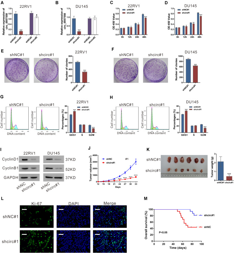
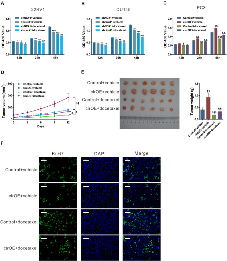
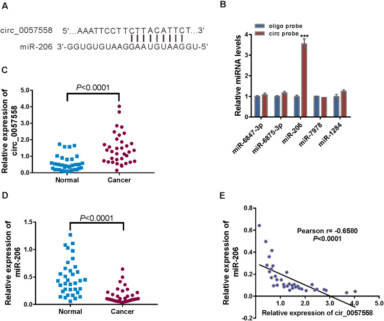
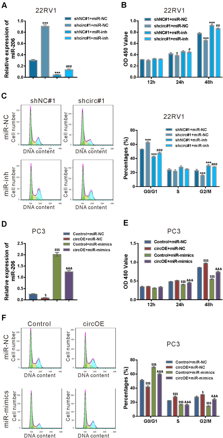
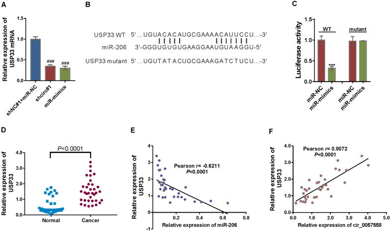
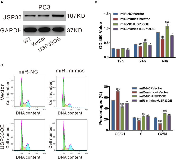
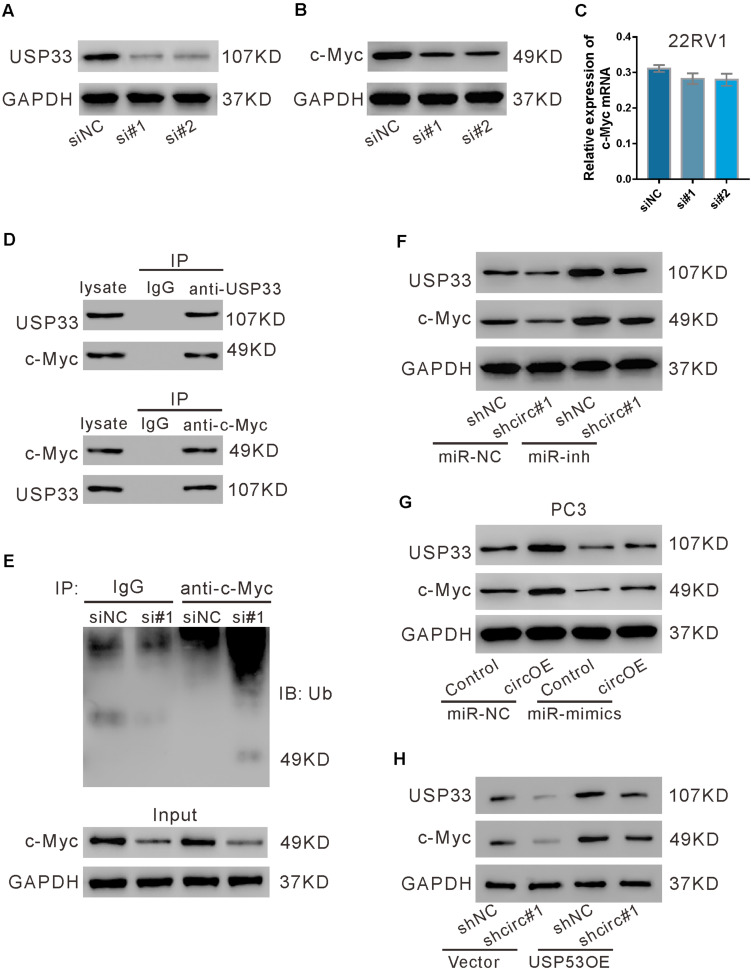
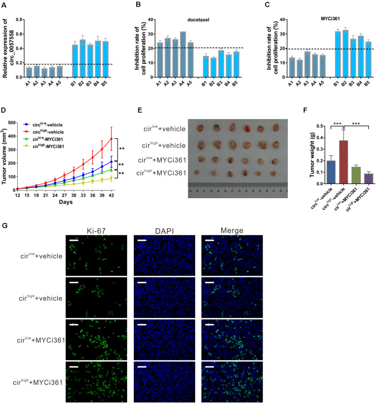
We previously reported the elevated expression of circ_0057558 in prostate cancer tissues and cell lines. Here, we aimed to determine the biological function of circ_0057558 in prostate cancer. In the current study, circ_0057558 knockdown in prostate cancer cells significantly repressed cell proliferation and colony formation, but promoted cell arrest and enhanced the sensitivity to docetaxel. Bioinformatics analysis prediction and RNA-pull down assay identified miR-206 as the potential binding miRNA of circ_0057558. A negative correlation was observed between the expression of miR-206 and circ_0057558 in prostate cancer tissues. miR-206 mimics rescued the function of circ_0057558 overexpression on prostate cancer cells. Further, the bioinformatics analysis and luciferase assay suggested that miR-206 may target ubiquitin-specific peptidase 33 (USP33). USP33 mRNA expression has negative correlation with miR-206 expression and positive correlation with circ_0057558 expression in prostate cancer tissues. USP33 overexpression partially blocked the effects of miR-206 mimics on prostate cell proliferation. USP33 could bind and deubiquitinate c-Myc. Increased c-Myc protein by circ_0057558 overexpression was partially reversed by miR-206 mimics. The proliferation inhibition activity of MYC inhibitor 361 (MYCi361) was more prominent in primary prostate cancer cells and patient-derived xenograft (PDX) model with higher level of circ_0057558. Collectively, circ_0057558 gives an impetus to cell proliferation and cell cycle control in prostate cancer cell lines by sponging miR-206 and positively regulating the transcription of the miR-206 target gene USP33.

Keywords: circular RNA; deubiquitination; proliferation; prostate cancer; sponge.


Conflict of interest statement

The authors declare that the research was conducted in the absence of any commercial or financial relationships that could be construed as a potential conflict of interest.

Figures

FIGURE 1
FIGURE 1
circ_0057558 knockdown suppressed prostate cancer cell proliferation in vitro and in vivo. (A,B) Lentivirus expressing specific shRNAs targeting circ_0057558 (shcirc#1 and shcir#2) and control shRNAs (shNC#1 and shNC#2) were infected into 22RV1 (A) and DU145 cells (B). circ_0057558 expression was detected by qRT-PCR at 48 h post infection. Wild-type cells (WT) that had no treatment were used as negative control. (C,D) CCK-8 assay was carried out to detect proliferation in circ_0057558 knockdown group (shcirc#1) and control group (shNC#1). (E,F) Colony formation assay was conducted to determine the colony-forming ability of circ_0057558 knockdown group (shcirc#1) and control group (shNC#1). (G,H) Cell cycle analysis detected by PI staining and flow cytometry analysis. (I) CyclinD1 and CyclinB1 were detected by western blotting. (J–M) 22RV1 cells infected with circ_0057558 shRNA (shcirc#1) or control shRNA (shNC#1) were transplanted into nude mice (n = 6 per group). The tumor growth curves (J), as well as the photos and weight (K) of xenografts on 33 days after inoculation are shown. Immunofluoresence staining with anti-Ki-67 (L) was carried out to assess cell proliferation in xenografts. Scale bar: 50 μm. (M) The Kaplan–Meier plot of survival duration in nude mice transplanted with 22RV1 inoculation infected with shcirc#1 or shNC#1 (n = 12 per group). Data were expressed as mean ± SD. *P < 0.05, **P < 0.01, ***P < 0.001 versus shNC#1; #P < 0.05 versus shNC#2.
FIGURE 2
FIGURE 2
circ_0057558 expression level influenced the sensitivity of prostate cancer cells to docetaxel. (A–C) 22RV1 (A) and DU145 cells (B) were infected with lentivirus expressing specific shRNAs targeting circ_0057558 (shcirc#1) and control shRNAs (shNC#1), while PC3 cells were transfected with plasmid expressing circ_0057558 (circOE)/control vector (control). These cell lines were then exposed to 10 nM docetaxel or vehicle (DMSO). Cell proliferation was measured with CCK-8 assay. (D–F) Nude mice were divided into four groups (n = 6 per group) and subcutaneously injected with PC3 cells stably expressing circ_0057558 (cirOE) or control vector (5 × 10⁶ cells per mouse, n = 12 per cells). When the volume of xenograft reached 100 mm3, the mice were intraperitoneally administered with docetaxel (10 mg/kg/day) or vehicle every 3 days. The tumor growth curves (D), as well as the photos and weight (E) of xenografts on 33 days after inoculation are shown. Immunofluoresence staining with anti-Ki-67 (F) was carried out to assess cell proliferation in xenografts. Scale bar: 50 μm. *P < 0.05, **P < 0.01, ***P < 0.001 versus shNC#1 + vehicle; #P < 0.05, ##P < 0.01, ###P < 0.001 versus shNC#1 + docetaxel; $P < 0.05, $$P < 0.01, $$$P < 0.001 versus control + vehicle; &P < 0.05, &&P < 0.01, &&&P < 0.001 versus control + docetaxel.
FIGURE 3
FIGURE 3
circ_0057558 was targeted by miR-206. (A) The putative binding sites of miR-206 and circ_0057558. (B) qRT-PCR analysis of the expression of candidate miRNAs in 22RV1 cells after biotin pull-down assay. ***P < 0.001 versus oligo probe. (C,D) Expression of circ_0057558 (C) and miR-206 (D) in prostate cancer tissue and adjacent non-tumor tissue. (E) Pearson r analysis showed the correlation between the expression of circ_0057558 and miR-206 in prostate cancer tissues.
FIGURE 4
FIGURE 4
miR-206 inhibitor rescued the function of circ_0057558 knockdown, while miR-206 mimics rescued the function of circ_0057558 overexpression. (A–C) 22RV1 cells were transduced with lentivirus expressing circ_0057558 shRNA (shcirc#1)/control shRNA (shNC#1) and transfected with miR-206 inhibitor (miR-inh)/control (miR-NC). miR-206 expression (A) was detected by qRT-PCR at 48 h post treatment. CCK-8 assay (B) and cell cycle analysis (C) was carried out to detect cell proliferation and cell cycle distribution, respectively. *P < 0.05, ***P < 0.001 versus shNC#1 + miR-NC; #P < 0.05, ##P < 0.01, ###P < 0.001 versus shcirc#1 + miR-NC. (D–F) PC3 cells were transfected with plasmid expressing circ_0057558 (circOE)/control vector (control) and transfected with miR-206 mimics (miR-mimics)/control (miR-NC). qRT-PCR (D), CCK-8 assay (E), and cell cycle analysis (F) were done to detect miR-206 expression, cell proliferation, and cell cycle distribution, respectively. $P < 0.05, $$$P < 0.001 versus control + miR-NC; &&&P < 0.001 versus circOE + miR-NC.
FIGURE 5
FIGURE 5
circ_0057558 regulated the miR-206 target, USP33. (A) mRNA expression of USP33 in 22RV cells infected with lentivirus expressing circ_0057558 shRNA (shcirc#1) or transfected with miR-206 mimics (miR-mimics). Cells infected with control shRNA (shNC#1) and transfected with control miRNA (miR-NC) were served as control. ###P < 0.001 versus shNC#1 + miR-NC. (B) The putative miRNA binding sites in the USP33 3′UTR. (C) pGL3-USP33 wild-type (WT) or mutant was co-transfected into 22RV1 cells with miR-206 mimics (miR-mimics) or control (miR-NC). Luciferase assay was performed to determine luciferase activity. The relative luciferase activity normalized to the control group (miR-NC). ***P < 0.001 versus miR-NC. (D) qRT-PCR analysis following the RIP assay was conducted to confirm the interaction between miR-206 with USP33. $$$P < 0.001 versus IgG. (E) Expression of USP33 mRNA in prostate cancer tissue and adjacent non-tumor tissue. (F,G) Pearson r analysis showed the correlation between the expression of miR-206 and USP33 and the expression of circ_0057558 and USP33 in prostate cancer tissues.
FIGURE 6
FIGURE 6
USP33 overexpression rescued the function of miR-206 mimics. (A) Protein expression of USP33 in PC3 cells infected with lentivirus expressing USP33 (USP33OE)/control vector (Vector). Representative blots from three independent experiments are shown. (B–D) PC3 cells were transfected with USP33OE/vector and transfected with miR-206 mimics (miR-mimics)/control (miR-NC). CCK-8 assay (B) and cell cycle analysis (C) were done to detect cell proliferation and cell cycle distribution, respectively. (D) CyclinD1 and CyclinB1 were detected by western blotting. **P < 0.01, ***P < 0.001 versus miR-NC + vector; #P < 0.05, ###P < 0.001 versus miR-mimics + vector.
FIGURE 7
FIGURE 7
USP33 deubiquitinated c-Myc. (A–C) 22RV1 cells were transfected with USP33 siRNAs (si#1 and si#2) or control siRNA (siNC). After 48 h, the protein expression of USP33 (A) and c-Myc (B) and the mRNA expression of c-Myc (C) were evaluated. (D) 22RV1 cell lysate was immunoprecipitated (IP) with anti-USP33 (upper) or anti-c-Myc (lower) or control IgG and then immunoblotted (IB) with anti-USP33 and anti-c-Myc. (E) 22RV1 cells were transfected with USP33 siRNA (si#1) or siNC. Immunoprecipitation experiment was performed with anti-c-Myc or control IgG and IB with anti-ubiquitin (Ub). (F) 22RV1 cells were transduced with lentivirus expressing circ_0057558 shRNA (shcirc#1)/control shRNA (shNC#1) and transfected with miR-206 inhibitor (miR-inh)/control (miR-NC). The expression of USP33 and c-Myc was detected at 48 h post treatment. (G) PC3 cells were transfected with plasmid expressing circ_0057558 (circOE)/control vector (control) and miR-206 mimics (miR-mimics)/control (miR-NC). The expression of USP33 and c-Myc was assessed at 48 h post treatment. (H) 22RV1 cells were transduced with shcirc#1/shNC#1 and USP33OE/vector. The expression of USP33 and c-Myc was detected at 48 h post treatment.
FIGURE 8
FIGURE 8
circ_0057558 expression level was correlated with the proliferation inhibition effect of c-Myc inhibitor MYCi361. (A–C) Primary prostate cancer cells were isolated as described in Materials and Methods section. circ_0057558 expression (A) in the primary prostate cancer cells was assessed by qRT-PCR. Primary cells were treated with 10 nM docetaxel (B), 6 μM MYCi361 (C), or vehicle (DMSO) for 48 h. Inhibition rate of cell proliferation (%) was determined using CCK-8 assay. (D–G) Patient-derived xenograft (PDX) model was established with fresh prostate cancer tissues and defined as circ_0057558high group and circ_0057558low group. The F2 PDXs were treated with MYCi361 (55 mg/kg/day) and vehicle (DMSO) twice a week by intraperitoneal injection. Tumor growth curves (D) and the photos and weight of PDX (E) are shown. (F) Immunofluoresence staining with anti-Ki-67 was carried out to assess cell proliferation in xenografts. Scale bar: 50 μm. **P < 0.01, ***P < 0.001.

References

    1. Anderson C., Catoe H., Werner R. (2006). MIR-206 regulates connexin43 expression during skeletal muscle development. Nucleic Acids Res. 34 5863–5871. 10.1093/nar/gkl743 - DOI - PMC - PubMed
    1. Chen L.-L. (2016). The biogenesis and emerging roles of circular RNAs. Nat. Rev. Mol. Cell Biol. 17:205. 10.1038/nrm.2015.32 - DOI - PubMed
    1. Chen Q., Jiao D., Yan L., Wu Y., Hu H., Song J., et al. (2015). Comprehensive gene and microRNA expression profiling reveals miR-206 inhibits MET in lung cancer metastasis. Mol. BioSyst. 11 2290–2302. 10.1039/c4mb00734d - DOI - PubMed
    1. Cocquerelle C., Mascrez B., Hetuin D., Bailleul B. (1993). Mis-splicing yields circular RNA molecules. FASEB J. 7 155–160. 10.1096/fasebj.7.1.7678559 - DOI - PubMed
    1. Dai C., Xie Y., Zhuang X., Yuan Z. (2018). MiR-206 inhibits epithelial ovarian cancer cells growth and invasion via blocking c-Met/AKT/mTOR signaling pathway. Biomed. Pharmacother. 104 763–770. 10.1016/j.biopha.2018.05.077 - DOI - PubMed

LinkOut - more resources