. 2021 Feb 25:9:648261.
doi: 10.3389/fcell.2021.648261. eCollection 2021.

Maternal Lead Exposure Impairs Offspring Learning and Memory via Decreased GLUT4 Membrane Translocation

Affiliations

Maternal Lead Exposure Impairs Offspring Learning and Memory via Decreased GLUT4 Membrane Translocation

Zai-Hua Zhao et al. Front Cell Dev Biol. .

Abstract

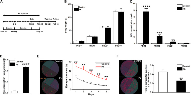
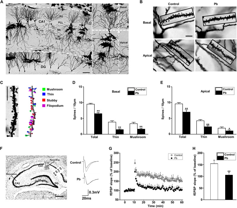
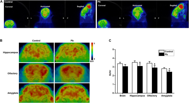
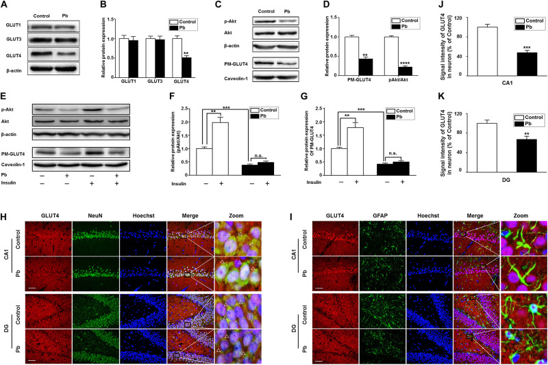
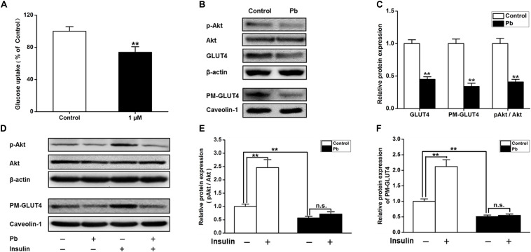
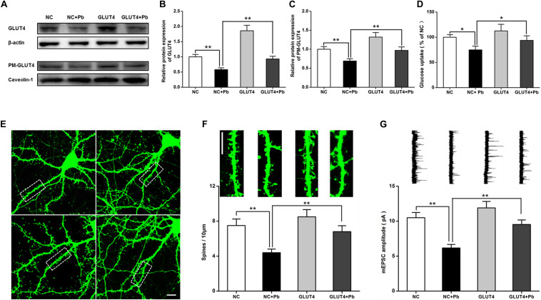
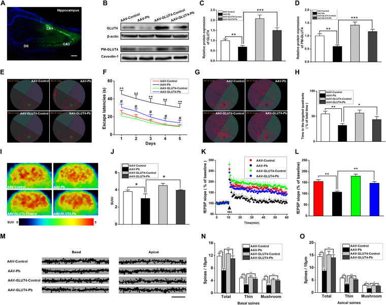
Lead (Pb) can cause a significant neurotoxicity in both adults and children, leading to the impairment to brain function. Pb exposure plays a key role in the impairment of learning and memory through synaptic neurotoxicity, resulting in the cognitive function. Researches have demonstrated that Pb exposure plays an important role in the etiology and pathogenesis of neurodegenerative diseases, such as Alzheimer's disease. However, the underlying mechanisms remain unclear. In the current study, a gestational Pb exposure (GLE) rat model was established to investigate the underlying mechanisms of Pb-induced cognitive impairment. We demonstrated that low-level gestational Pb exposure impaired spatial learning and memory as well as hippocampal synaptic plasticity at postnatal day 30 (PND 30) when the blood concentration of Pb had already recovered to normal levels. Pb exposure induced a decrease in hippocampal glucose metabolism by reducing glucose transporter 4 (GLUT4) levels in the cell membrane through the phosphatidylinositol 3 kinase-protein kinase B (PI3K-Akt) pathway. In vivo and in vitro GLUT4 over-expression increased the membrane translocation of GLUT4 and glucose uptake, and reversed the Pb-induced impairment to synaptic plasticity and cognition. These findings indicate that Pb exposure impairs synaptic plasticity by reducing the level of GLUT4 in the cell membrane as well as glucose uptake via the PI3K-Akt signaling pathway, demonstrating a novel mechanism for Pb exposure-induced neurotoxicity.

Keywords: PI3K-Akt; glucose transporter 4; hippocampus synaptic plasticity; lead; learning and memory.


Conflict of interest statement

The authors declare that the research was conducted in the absence of any commercial or financial relationships that could be construed as a potential conflict of interest.

Figures

FIGURE 1
FIGURE 1
Accumulation of Pb in the hippocampus of PND30 rats and its influence on learning and memory. (A) Establishment of Pb exposure model. Female SD rats were given Pb acetate solutions to drink (0 or 0.02%) beginning 2 weeks before mating until PND 10. (B) Body weights and (C) Blood Pb levels. The concentration of blood Pb in the experimental rats was measured at PND 0, 10, 21, and 30. (D) Pb levels in the hippocampus region. (E,F) Analysis of MWM results. The MWM tests were only measured at PND30. Data are expressed as mean ± SE, **p < 0.01, ***p < 0.001, ****p < 0.0001; n = 6 per group.
FIGURE 2
FIGURE 2
Hippocampal Pb accumulation and impairment of synaptic plasticity in PND30 rats. (A,B) Golgi Staining for basal and apical dendritic spines in hippocampal CA1 region. Scale bars = 100 μm in (A) and 10 μm in (B). (C) Imaris reconstruction of the dendritic spines of pyramidal neurons in CA1 region. Scale bar = 10 μm. (D,E) Statistical analysis of the effect of Pb exposure on basal and apical spines. For each group, data are expressed as mean ± SE of spines per 10 μm (16 neurons/8 rats for all groups), *p < 0.05, **p < 0.01. (F) LTP induced by high-frequency tetanic stimulation. Simulations were applied to the CA3 regions of the hippocampus and the signal was collected at the CA1 region. Scale bar = 500 μm. (G,H) LTP in hippocampal regions CA3-CA1. Abscissa represented the time of tetanic stimulation. N = 16 slices per 8 rats in each group. The patch-clamp recordings showed that the fEPSP slope for rat hippocampal LTP in the Pb-exposed group was significantly reduced compared with that in the control group. Data are expressed as mean ± SE, **p < 0.01. SO, stratum oriens; PCL, pyramidal cell layer; SR, stratum radiatum; SL-M, stratum lacunosum-moleculare; DG, dentate gyrus.
FIGURE 3
FIGURE 3
Effect of low-level gestational Pb exposure on glucose uptake in rat brain. (A) Representative images of experimental rat brains. (B) Representative images of glucose uptake in different brain areas ([18F]-FDG microPET/CT) in the experimental rats at the last scanning point. (C) Statistical analysis of PET-CT scanning data. PET-CT results indicated that maternal Pb exposure at a low level significantly reduced the level of glucose metabolism in the rat hippocampus. *p < 0.05, n = 6 per group.
FIGURE 4
FIGURE 4
Effects of Pb exposure on GLUT4 expression and membrane translocation in rat hippocampus. (A–D) Effects of Pb exposure on the expression of GLUTs, PM-GLUT4, and p-Akt. There were no remarkable changes in GLUT3 and GLUT1 expression in Pb-exposed rat hippocampus, but the GLUT4 expression was significantly reduced. The expression levels of PM-GLUT4 and p-Akt were significantly reduced. Among them, caveolin-1 served as the loading control of PM-GLUT4, and β-actin for other proteins. (E–G) Effects of insulin on PM-GLUT4 and p-Akt expression levels. Insulin stimulation significantly increased the expression of p-Akt in rat hippocampus but did not reverse the decrease of p-Akt expression in the Pb-exposed group. N = 6 per group, **p < 0.01, ***p < 0.001, ****p < 0.0001. vs. control. The band intensity was statistically analyzed based on the percentage relative to the control. (H,I) Immunofluorescence staining. (J,K) Statistical results of signal intensity of GLUT4 expression in neuron in CA1 and DG regions of the hippocampus in rats. The results showed that GLUT4 and NeuN were significantly double-labeled in the CA1 and DG regions of the hippocampus in rats, and double-labeling in the Pb-exposed group was significantly reduced compared to the control group levels. Immunofluorescence staining indicated that GLUT4 and GFAP were not double-labeled in the hippocampal CA1 and DG regions of rats. Scale bars = 50 μm. Data were represented as mean ± SEM and counted based on data from a minimum of three independent experiments, **p < 0.01, ***p < 0.001.
FIGURE 5
FIGURE 5
Effects on the glucose uptake and GLUT4, PM-GLUT4, and p-Akt expression in primary hippocampal neurons in the Pb-exposed rats. (A) Effect of Pb exposure on the glucose uptake. Using 2-NBDG combined with flow cytometry, glucose uptake in primary cultured rat hippocampal neurons was analyzed. Flow cytometry results showed that glucose uptake was significantly reduced in primary cultured hippocampal neurons after treatment with 1 μmol/L Pb acetate compared with the levels in the control group. (B,C) Effect of Pb exposure on expression of glycometabolism-related proteins in the brain. The expression levels of GLUT4, PM-GLUT4, and pAkt proteins in primary cultured hippocampal neurons were dramatically reduced in the Pb-exposed rats. (D–F) p-Akt and PM-GLUT4 expression after insulin stimulation in rat primary cultured hippocampal neurons. Insulin stimulation led to the significant increase of p-Akt and PM-GLUT4 protein levels in the control rat hippocampus, but did not reverse the decreased levels in Pb-exposed rats. Among them, caveolin-1 served as the loading control of PM-GLUT4, and β-actin for other proteins. **p < 0.01, n = 6 per group. Statistical analysis of the band intensity measured as a percentage of the control.
FIGURE 6
FIGURE 6
Effects of over-expression of GLUT4 on glucose uptake and synaptic plasticity of primary hippocampal neurons in the Pb-exposed rats. (A–C) Expression changes in GLUT4 and PM-GLUT4 in rat hippocampal neurons after AAV-GLUT4 transfection. GLUT4 and PM-GLUT4 expression levels returned to normal in the rat primary cultured hippocampal neurons of the Pb-exposed group after AAV-GLUT4 transfection. Among them, caveolin-1 served as the loading control of PM-GLUT4, and β-actin for GLUT4. (D) Effect of GLUT4 over-expression on glucose uptake in rat hippocampal neurons. Over-expression of GLUT4 recovered the glucose uptake levels in the primary hippocampal neurons from the Pb-exposed group. (E–G) Effects of GLUT4 over-expression on cell morphology and LTP in Pb-exposed rat hippocampal neurons. After lentivirus infection, the morphology (dendritic spines) of rat primary cultured hippocampal neurons was observed via laser confocal microscopy. Over-expression of GLUT4 reversed the dendritic spine density and LTP lesions in Pb-exposed rats. Scale bar = 10 μm. *p < 0.05, **p < 0.01, vs. control, n = 6 per group.
FIGURE 7
FIGURE 7
Effects of GLUT4 over-expression in Pb-exposed rat hippocampus on the glucose uptake, synaptic plasticity, as well as learning and memory function. (A–D) Over-expression of GLUT4 in rat hippocampus and analysis of its effect on glycometabolism-related proteins. GLUT4 over-expression was induced via stereotactic injection of adenovirus. GLUT4 over-expression reversed the decreased PM-GLUT4 in Pb-exposed rat hippocampus. (E–H) Analysis of rat positioning, navigation ability, and space exploration trajectory. Over-expression of GLUT4 in Pb-exposed rat hippocampus reversed the arrival latency and the target quadrant retention time of model rats in MWM tests; *p < 0.05, **p < 0.01; # p < 0.05 vs. AAV-Pb. (I,J) PET-CT scanning and analysis of glucose metabolism in rat brain. The glucose metabolism level in Pb-exposed rat hippocampus was reversed after the over-expression of GLUT4. (K,L) Effects of Pb exposure on LTP induction in rat hippocampus. After over-expression of GLUT4 in rat hippocampus, the slope of fEPSP in Pb-exposed rat hippocampus was reversed. (M–O) Effect of Pb exposure on dendritic spines of hippocampal pyramidal neurons. Over-expression of GLUT4 increased the spinal density of basal and apical dendrites in pyramidal neurons as well as the number of thin and mushroom-shaped dendritic spines in the Pb-exposed rat hippocampus. Scale bar = 10 μm. *p < 0.05, **p < 0.01, ***p < 0.001, vs. control, n = 6 per group.

Similar articles

Cited by

References

    1. Ahmad F., Salahuddin M., Alamoudi W., Acharya S. (2018). Dysfunction of cortical synapse-specific mitochondria in developing rats exposed to lead and its amelioration by ascorbate supplementation. Neuropsychiatr. Dis. Treat. 14 813–824. 10.2147/NDT.S148248 - DOI - PMC - PubMed
    1. Anderson D. W., Mettil W. A., Schneider J. S. (2013). Rearing environment, sex and developmental lead exposure modify gene expression in the hippocampus of behaviorally naive animals. Neurochem. Int. 62 510–520. 10.1016/j.neuint.2013.01.003 - DOI - PMC - PubMed
    1. Barha C. K., Galea L. A. (2013). The hormone therapy, premarin, impairs hippocampus-dependent spatial learning and memory and reduces activation of new granule neurons in response to memory in female rats. Neurobiol. Aging 34 986–1004. 10.1016/j.neurobiolaging.2012.07.009 - DOI - PubMed
    1. Bhattacharya A., Shukla R., Auyang E. D., Dietrich K. N., Bornschein R. (2007). Effect of succimer chelation therapy on postural balance and gait outcomes in children with early exposure to environmental lead. Neurotoxicology 28 686–695. 10.1016/j.neuro.2007.03.007 - DOI - PubMed
    1. Bihaqi S. W., Zawia N. H. (2013). Enhanced taupathy and AD-like pathology in aged primate brains decades after infantile exposure to lead (Pb). Neurotoxicology 39 95–101. 10.1016/j.neuro.2013.07.010 - DOI - PMC - PubMed

LinkOut - more resources