

Relationship of Anterior Cruciate Ligament Volume and T2* Relaxation Time to Anterior Knee Laxity
- PMID: 33718498
- PMCID: PMC7925955
- DOI: 10.1177/2325967120979986
Relationship of Anterior Cruciate Ligament Volume and T2* Relaxation Time to Anterior Knee Laxity
Abstract
Background: High anterior knee laxity (AKL) has been prospectively identified as a risk factor for anterior cruciate ligament (ACL) injuries. Given that ACL morphometry and structural composition have the potential to influence ligamentous strength, understanding how these factors are associated with greater AKL is warranted.
Hypothesis: Smaller ACL volumes combined with longer T2* relaxation times would collectively predict greater AKL.
Study design: Cross-sectional study; Level of evidence, 3.
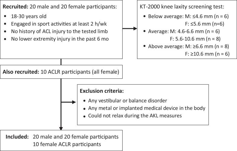
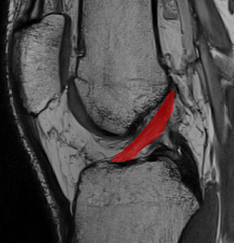
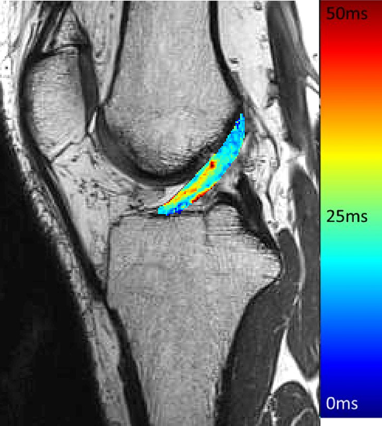
Methods: College-aged active male (n = 20) and female (n = 30) participants underwent magnetic resonance imaging (MRI) and AKL testing. T2-weighted MRI scans were used to assess ACL volumes, and T2* relaxation times were used to assess ACL structural composition. AKL was measured via a commercial knee arthrometer. Forward stepwise linear regression with sex and weight (first step; suppressor variables) as well as ACL volume and T2* relaxation time (second step; independent variables) was used to predict AKL (dependent variable).
Results: After initially adjusting for sex and weight (R 2 = 0.19; P = .006), smaller ACL volumes combined with longer T2* relaxation times collectively predicted greater AKL (R 2 = 0.52; P < .001; R 2 Δ = 0.32; P Δ < .001). A smaller ACL volume was the primary predictor of greater AKL (R 2 Δ = 0.28; P < .001), with a longer T2* relaxation time trending toward a significant contribution to greater AKL (R 2 Δ = 0.04; P = .062). After adjusting for ACL volume and T2* relaxation time, sex (partial r = 0.05; P = .735) and weight (partial r = 0.05; P = .725) were no longer significant predictors.
Conclusion: AKL was largely predicted by ACL volume and to a lesser extent by T2* relaxation time (and not a person's sex and weight). These findings enhance our understanding of how AKL may be associated with a structurally weaker ACL. The current study presents initial evidence that AKL is a cost-effective and clinically accessible measure that shows us something about the structural composition of the ACL. As AKL has been consistently shown to be a risk factor for ACL injuries, work should be done to continue to investigate what AKL may tell a clinician about the structure and composition of the ACL.
Keywords: ACL size; MRI; knee; relaxation times.
© The Author(s) 2021.
Conflict of interest statement
One or more of the authors has declared the following potential conflict of interest or source of funding: This work was supported by the Gateway MRI Center at the University of North Carolina at Greensboro. AOSSM checks author disclosures against the Open Payments Database (OPD). AOSSM has not conducted an independent investigation on the OPD and disclaims any liability or responsibility relating thereto.
Figures
References
-
- Amiel D, Ishizue KK, Harwood FL, Kitabayashi L, Akeson WH. Injury of the anterior cruciate ligament: the role of collagenase in ligament degeneration. J Orthop Res. 1989;7(4):486–493. - PubMed
-
- Anderson AF, Dome DC, Gautam S, Awh MH, Rennirt GW. Correlation of anthropometric measurements, strength, anterior cruciate ligament size, and intercondylar notch characteristics to sex differences in anterior cruciate ligament tear rates. Am J Sports Med. 2001;29(1):58–66. - PubMed
-
- Beynnon BD, Johnson RJ, Toyama H, et al. The relationship between anterior-posterior knee laxity and the structural properties of the patellar tendon graft: a study in canines. Am J Sports Med. 1994;22(6):812–820. - PubMed
LinkOut - more resources
Full Text Sources
Other Literature Sources
Research Materials
