

Reverse contralateral proximal tibial plating and cannulated screws fixation for Hoffa fracture: A case report
- PMID: 33718567
- PMCID: PMC7920851
- DOI: 10.1016/j.tcr.2021.100443
Reverse contralateral proximal tibial plating and cannulated screws fixation for Hoffa fracture: A case report
Erratum in
-
Erratum regarding missing Declaration of Competing Interest statements in previously published articles.Trauma Case Rep. 2023 Feb 17;45:100794. doi: 10.1016/j.tcr.2023.100794. eCollection 2023 Jun. Trauma Case Rep. 2023. PMID: 37234575 Free PMC article.
Abstract
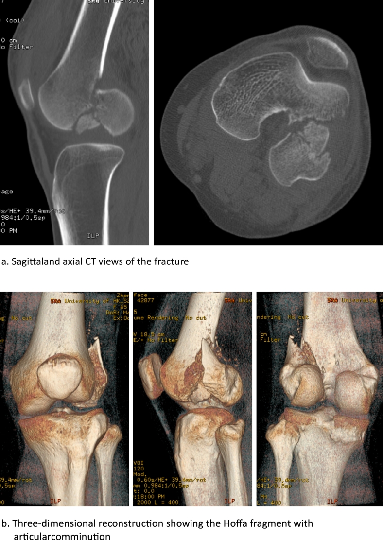
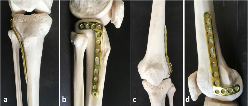
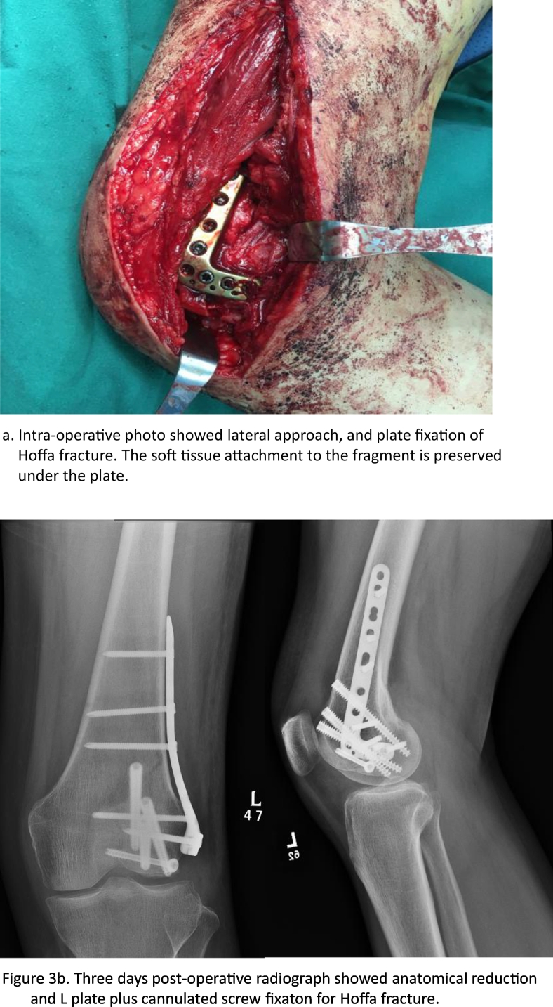
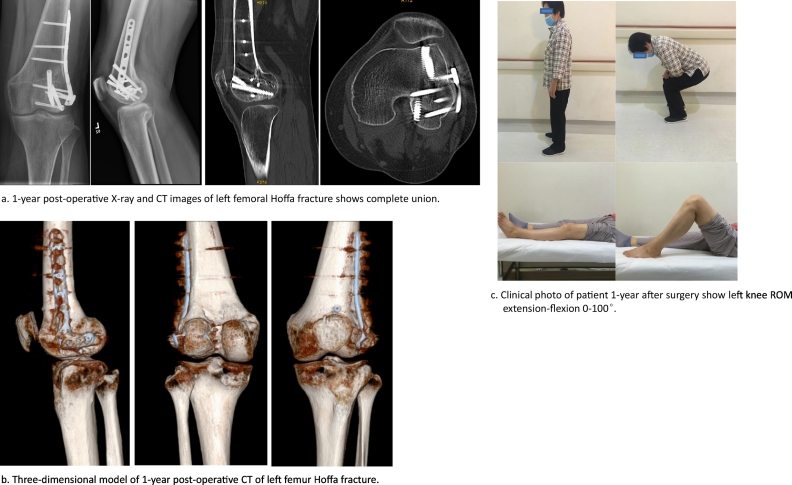
Hoffa fracture is a rare type of distal femoral fracture occurs in the coronal plane of either femoral epicondyle. To date, screws in combination with lateral plate fixation is widely accepted to achieve stable fixation and good results. However, up to now there has not been a specially designed anatomical plate for lateral fixation of Hoffa fracture. In this report, we demonstrate a case of Hoffa fracture fixed with reverse application of "L" shaped contralateral proximal tibia plate and cannulated screws, resulting in good one-year results.
Keywords: Hoffa fracture; Internal fixation; Open reduction; Reverse contralateral proximal tibia plate.
© 2021 The Authors.
Figures
References
-
- Lin T. Cannulated lag screw combined with lateral supporting plate for treatment of Hoffa fracture of Letenneur type I and type III. Zhongguo Xiu Fu Chong Jian Wai Ke Za Zhi. 2013;27(9):1050–1053. - PubMed
-
- Zhao L.L., Tong P.J., Xiao L.W. Internal fixation with lag screws plus an anti-sliding plate for the treatment of Hoffa fracture of the lateral femoral condyle. Zhongguo Gu Shang. 2016;29(3):266–269. - PubMed
-
- Bartonicek J., Rammelt S. History of femoral head fracture and coronal fracture of the femoral condyles. Int. Orthop. 2015;39(6):1245–1250. - PubMed
Publication types
LinkOut - more resources
Full Text Sources
Other Literature Sources
