

The application of optical coherence tomography angiography in Alzheimer's disease: A systematic review
- PMID: 33718582
- PMCID: PMC7927164
- DOI: 10.1002/dad2.12149
The application of optical coherence tomography angiography in Alzheimer's disease: A systematic review
Abstract
Introduction: Discovering non-invasive and easily acquired biomarkers that are conducive to the accurate diagnosis of dementia is an urgent area of ongoing clinical research. One promising approach is retinal imaging, as there is homology between retinal and cerebral vasculature. Recently, optical coherence tomography angiography (OCT-A) has emerged as a promising new technology for imaging the microvasculature of the retina.
Methods: A systematic review and meta-analysis was conducted to examine the application of OCT-A in dementia.
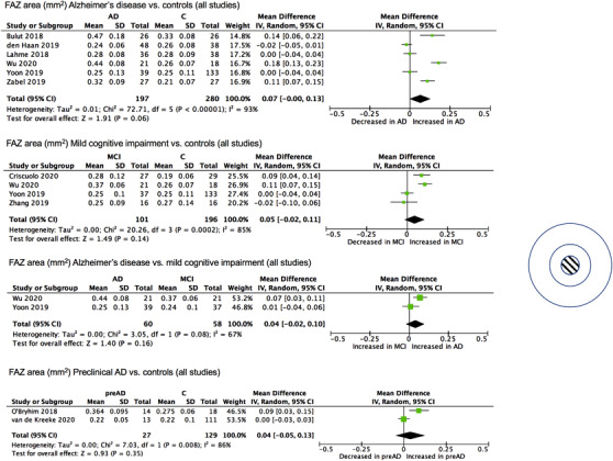
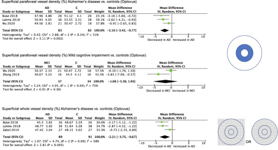
Results: Fourteen studies assessing OCT-A in preclinical Alzheimer's disease (AD), mild cognitive impairment, or AD were included. Exploratory meta-analyses revealed a significant increase in the foveal avascular zone area and a significant decrease in superficial parafoveal and whole vessel density in AD, although there was significant heterogeneity between studies.
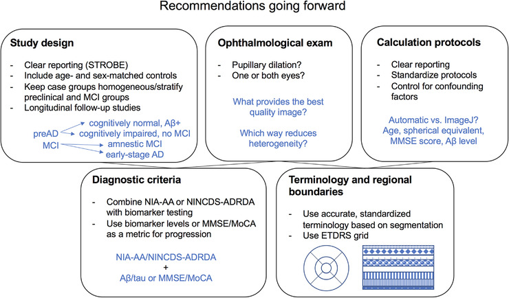
Discussion: Although certain OCT-A metrics may have the potential to serve as biomarkers for AD, the field requires further standardization to allow conclusions to be reached regarding their clinical utility.
Keywords: Alzheimer's disease; dementia; diagnostic tool; foveal avascular zone; mild cognitive impairment; optical coherence tomography angiography; perfusion density; preclinical; retinal imaging; retinal vasculature; vessel density.
© 2021 The Authors. Alzheimer's & Dementia: Diagnosis, Assessment & Disease Monitoring published by Wiley Periodicals, LLC on behalf of Alzheimer's Association.
Conflict of interest statement
The authors do not have any conflicts of interest.
Figures
Similar articles
-
Retinal imaging biomarkers of Alzheimer's disease: A systematic review and meta-analysis of studies using brain amyloid beta status for case definition.Alzheimers Dement (Amst). 2023 May 25;15(2):e12421. doi: 10.1002/dad2.12421. eCollection 2023 Apr-Jun. Alzheimers Dement (Amst). 2023. PMID: 37250908 Free PMC article. Review.
-
Retinal vascular biomarkers in mild cognitive impairment and Alzheimer's disease: a comprehensive review and meta-analysis.Alzheimers Dement (Amst). 2025 Jun 16;17(2):e70132. doi: 10.1002/dad2.70132. eCollection 2025 Apr-Jun. Alzheimers Dement (Amst). 2025. PMID: 40525098 Free PMC article. Review.
-
Unlocking the Potential of Vessel Density and the Foveal Avascular Zone in Optical Coherence Tomography Angiography as Biomarkers in Alzheimer's Disease.Healthcare (Basel). 2024 Aug 9;12(16):1589. doi: 10.3390/healthcare12161589. Healthcare (Basel). 2024. PMID: 39201148 Free PMC article. Review.
-
Optical coherence tomography angiography measurements in Parkinson's disease: A systematic review and meta-analysis.Eye (Lond). 2023 Oct;37(15):3145-3156. doi: 10.1038/s41433-023-02483-2. Epub 2023 Mar 20. Eye (Lond). 2023. PMID: 36941403 Free PMC article.
-
Retinal microvascular attenuation in mental cognitive impairment and Alzheimer's disease by optical coherence tomography angiography.Acta Ophthalmol. 2020 Sep;98(6):e781-e787. doi: 10.1111/aos.14381. Epub 2020 Mar 9. Acta Ophthalmol. 2020. PMID: 32153141
Cited by
-
Optical coherence tomography measurements in Huntington's disease: a systematic review and meta-analysis.J Neurol. 2024 Oct;271(10):6471-6484. doi: 10.1007/s00415-024-12634-4. Epub 2024 Aug 26. J Neurol. 2024. PMID: 39187741 Free PMC article.
-
Detection of Retinal Microvascular Changes with Optical Coherence Tomography Angiography in Patients with Acute Leukemia Without Retinopathy.Ophthalmol Ther. 2024 May;13(5):1145-1157. doi: 10.1007/s40123-024-00904-3. Epub 2024 Feb 28. Ophthalmol Ther. 2024. PMID: 38416329 Free PMC article.
-
Retinal imaging biomarkers of Alzheimer's disease: A systematic review and meta-analysis of studies using brain amyloid beta status for case definition.Alzheimers Dement (Amst). 2023 May 25;15(2):e12421. doi: 10.1002/dad2.12421. eCollection 2023 Apr-Jun. Alzheimers Dement (Amst). 2023. PMID: 37250908 Free PMC article. Review.
-
Using Optical Coherence Tomography to Screen for Cognitive Impairment and Dementia.J Alzheimers Dis. 2021;84(2):723-736. doi: 10.3233/JAD-210328. J Alzheimers Dis. 2021. PMID: 34569948 Free PMC article.
-
Automated Foveal Avascular Zone Segmentation in Optical Coherence Tomography Angiography Across Multiple Eye Diseases Using Knowledge Distillation.Bioengineering (Basel). 2025 Mar 23;12(4):334. doi: 10.3390/bioengineering12040334. Bioengineering (Basel). 2025. PMID: 40281694 Free PMC article.
References
Publication types
Grants and funding
LinkOut - more resources
Full Text Sources
Other Literature Sources