. 2021 Feb 4:20:447-458.
doi: 10.1016/j.omto.2021.01.017. eCollection 2021 Mar 26.

Applications of nuclear-based imaging in gene and cell therapy: probe considerations

Affiliations

Applications of nuclear-based imaging in gene and cell therapy: probe considerations

Alessia Volpe et al. Mol Ther Oncolytics. .

Abstract

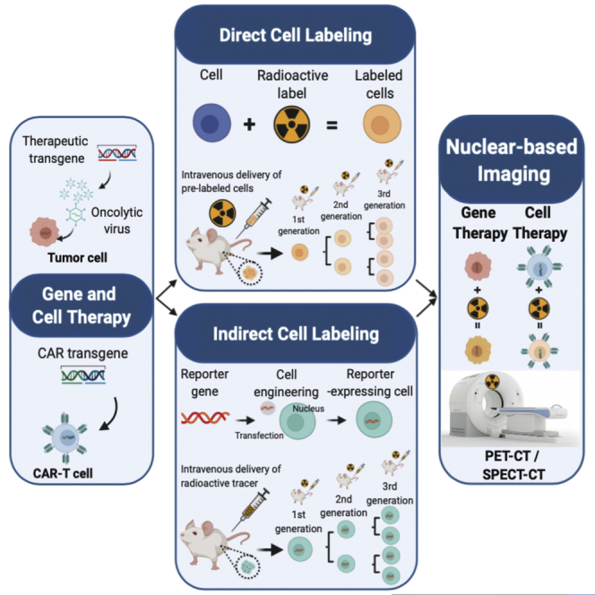
Several types of gene- and cell-based therapeutics are now emerging in the cancer immunotherapy, transplantation, and regenerative medicine landscapes. Radionuclear-based imaging can be used as a molecular imaging tool for repetitive and non-invasive visualization as well as in vivo monitoring of therapy success. In this review, we discuss the principles of nuclear-based imaging and provide a comprehensive overview of its application in gene and cell therapy. This review aims to inform investigators in the biomedical field as well as clinicians on the state of the art of nuclear imaging, from probe design to available radiopharmaceuticals and advances of direct (probe-based) and indirect (transgene-based) strategies in both preclinical and clinical settings. Notably, as the nuclear-based imaging toolbox is continuously expanding, it will be increasingly incorporated into the clinical setting where the distribution, targeting, and persistence of a new generation of therapeutics can be imaged and ultimately guide therapeutic decisions.

Keywords: cell therapy; gene therapy; nuclear imaging; radiotracer.


Conflict of interest statement

The authors declare no competing interests.

Figures

None
Graphical abstract
Figure 1
Figure 1
Application of preclinical direct nuclear imaging in cell therapy (A) CT, SPECT, and SPECT-CT images obtained at 2, 24, 48, and 120 h after 111In-oxine-labeled hemagglutinin (HA)-specific CTL administration. CT44 HA+ and CD26 HA colon tumors were established in the right and left footpad, respectively. Directly labeled cells using 89Zr-oxine in rhesus macaques is shown. (B) PET-CT of autologous 89Zr-oxine-labeled NK cells was performed after infusion and treatment with deferoxamine. Adoptive NK cell trafficking was monitored for up to 7 days and showed initial localization to the lungs, followed by a progressive distribution in liver and spleen. Bladder uptake is due to renal excretion of free 89Zr being released from dead/dying cells and chelated by deferoxamine. All figures adapted with permission from publishers.
Figure 3
Figure 3
Clinical application of radionuclide-based imaging using indirect and direct strategies (A) HSV1-tk reporter paired with [18F]FHBG PET radiotracer was successfully used for the non-invasive detection of cytolytic CAR-T cells in patients with recurrent glioma. T1W-weighted (T1W) MRI was used to assess tumor extent before and after intratumoral CAR-T cell administration (top), and images were superimposed with [18F]FHBG PET (bottom). (B) Dynamic SPECT showing the distribution of 99mTc-HMPAO-labeled neutrophils and eosinophils in a healthy volunteer. Accumulation of the two cell types appears to be different over time and mainly in lungs, liver, and spleen. All figures adapted with permission from publishers.
Figure 2
Figure 2
Application of preclinical reporter-based nuclear imaging in cell therapy Preclinical imaging of cell-based immunotherapies with multiple host-compatible reporter genes and their corresponding radioisotopes. (A) NIS-based 99 mTcO4 imaging of AdlP1 (top) and AdAM6 (bottom) adenoviruses in a HCT116 colorectal carcinoma xenograft. T, tumor; S, stomach. (B) Dual-modality PET-SPECT imaging with [123I]MIBG and [124I]FIAU to visualize intratumorally injected hNET-CD4+ and HSV1-tk-CD8+ T cell subpopulations in a human EBV lymphoma xenograft. (C) NIS-based [18F]BF4-afforded PET reveals CAR T cell retention differences in two models of TNBC. Endogenous NIS expression, prevalently in thyroid and stomach, does not interfere with imaging. ThSG, thyroid+salivary gland; S, stomach; yellow arrows indicate CAR T cells homing at the tumor. (D) High accumulation of dCKDM/GFP-expressing T cells is observed at the tumor site of treated mice through [18F]FEAU PET imaging. The region of interest is marked by the arrow. (E) Longitudinal PET-CT scan of SSTR2-expressing CAR T cells using 68Ga-DOTATOC. CAR T cells were administered. Indicated images are from the lungs at day 20 after tumor establishment. L, lungs. All figures adapted with permission from publishers.

Similar articles

Cited by

References

    1. Blasberg R.G., Tjuvajev J.G. Molecular-genetic imaging: current and future perspectives. J. Clin. Invest. 2003;111:1620–1629. - PMC - PubMed
    1. Peñuelas I., Haberkorn U., Yaghoubi S., Gambhir S.S. Gene therapy imaging in patients for oncological applications. Eur. J. Nucl. Med. Mol. Imaging. 2005;32(Suppl 2):S384–S403. - PubMed
    1. Vaquero J.J., Kinahan P. Positron emission tomography: current challenges and opportunities for technological advances in clinical and preclinical imaging systems. Annu. Rev. Biomed. Eng. 2015;17:385–414. - PMC - PubMed
    1. Khalil M.M., Tremoleda J.L., Bayomy T.B., Gsell W. Molecular SPECT imaging: an overview. Int. J. Mol. Imaging. 2011;2011:796025. - PMC - PubMed
    1. Basu S., Kwee T.C., Surti S., Akin E.A., Yoo D., Alavi A. Fundamentals of PET and PET/CT imaging. Ann. N Y Acad. Sci. 2011;1228:1–18. - PubMed

LinkOut - more resources