. 2021 Feb 6:20:484-498.
doi: 10.1016/j.omto.2021.02.005. eCollection 2021 Mar 26.

M2 macrophage-derived exosomal microRNA-155-5p promotes the immune escape of colon cancer by downregulating ZC3H12B

Affiliations

M2 macrophage-derived exosomal microRNA-155-5p promotes the immune escape of colon cancer by downregulating ZC3H12B

Yu-Shui Ma et al. Mol Ther Oncolytics. .

Abstract

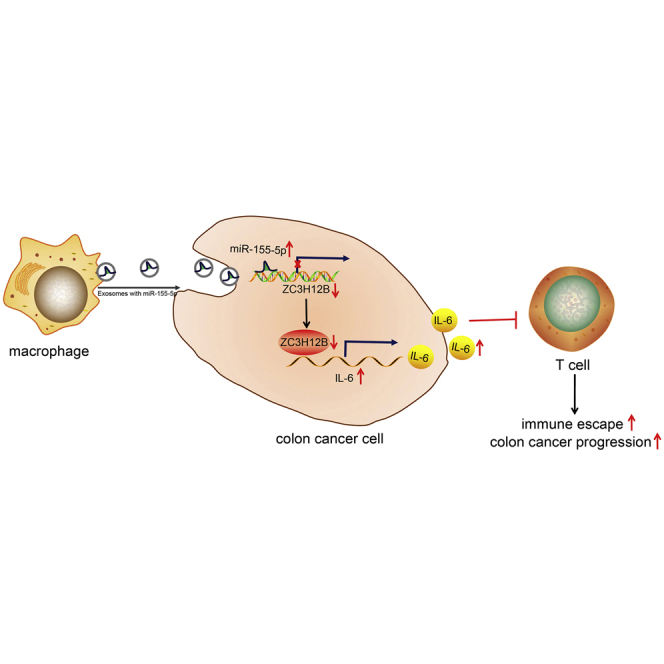
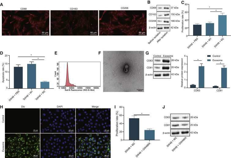
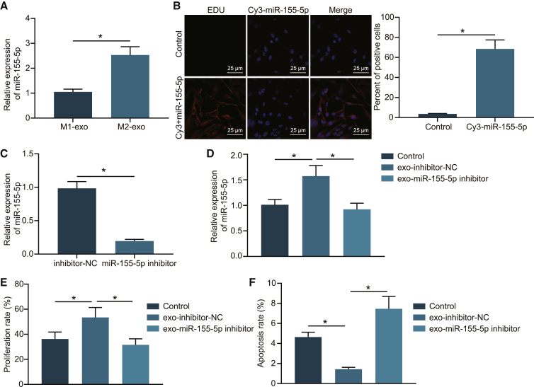
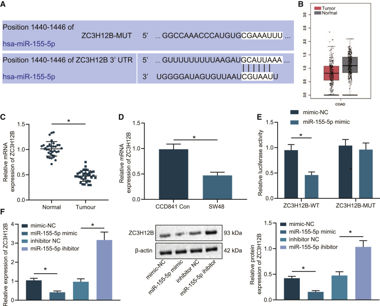
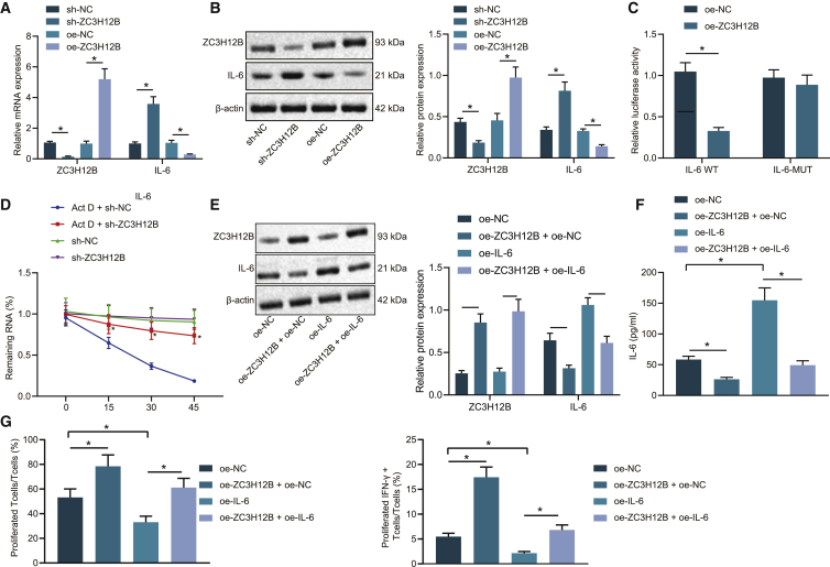
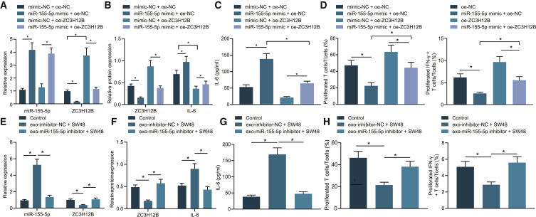
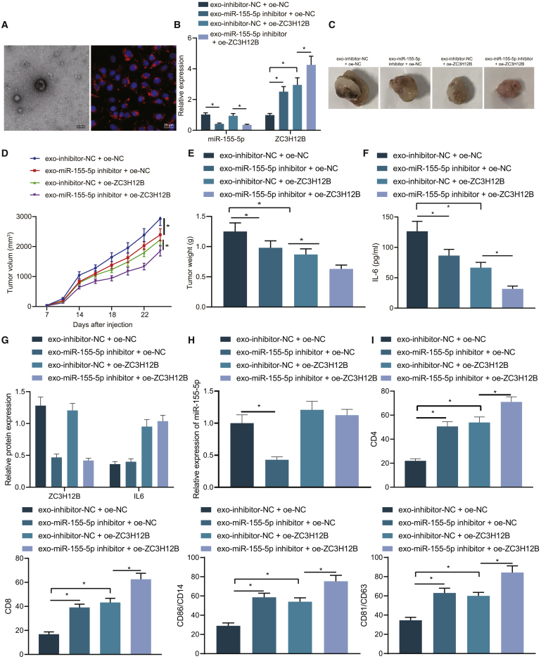
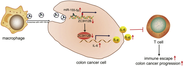
Previous evidence has highlighted M2 macrophage regulation of cancer cells via exosome shuttling of microRNAs (miRNAs or miRs). The current study set out to explore the possible role of M2 macrophage-derived exosomal miR-155-5p in regard to immune escape of colon cancer cells. Experimental data from quantitative reverse-transcriptase PCR (qRT-PCR) and western blot analysis revealed highly expressed miR-155-5p and interleukin (IL)-6 and poorly expressed ZC3H12B in M2 macrophage-derived exosomes. Additionally, miR-155-5p could be transferred by M2 macrophage-isolated exosomes to colon cancer cells, which targeted ZC3H12B by binding to the 3¢ UTR, as identified by dual luciferase reporter gene. Meanwhile, gain- and loss-of function experimentation on miR-155-5p and ZC3H12B in SW48 and HT29 cells cocultured with M2 macrophage-secreted exosomes demonstrated that miR-155-5p overexpression or ZC3H12B silencing promoted the proliferation and antiapoptosis ability of SW48 and HT29 cells, as well as augmenting the CD3+ T cell proliferation and the proportion of interferon (IFN)-γ+ T cells. Xenograft models confirmed that M2 macrophage-derived exosomal miR-155-5p reduced the ZC3H12B expression to upregulate IL-6, which consequently induced immune escape and tumor formation. Collectively, our findings indicated that M2 macrophage-derived exosomal miR-155-5p can potentially promote the immune escape of colon cancer by impairing ZC3H12B-mediated IL-6 stability reduction, thereby promoting the occurrence and development of colon cancer.

Keywords: M2 macrophages; ZC3H12B; colon cancer; exosomes; immune escape; microRNA-155-5p.


Conflict of interest statement

The authors declare no competing interests.

Figures

None
Graphical abstract
Figure 1
Figure 1
Colon cancer progression was affected by M2 macrophage-derived exosomes (A) Immunostaining of expression of M2 macrophage markers (CD68, CD163, and CD206) (×200). (B) Western blot analysis of protein expression patterns of the M2 macrophage markers (CD68, CD163, and CD206). (C) The effect of M2 macrophages on SW48 cell proliferation detected by EdU assay. (D) The effect of M2 macrophages on SW48 cell apoptosis rate measured by flow cytometry. (E) Nanoparticle tracking analysis of particle size of exosomes. (F) The ultrastructure of the exosomes (×5,000) observed under a transmission electron microscope. (G) Western blot analysis of the expression of exosome markers (CD63 and CD81). Control, the supernatant after exosome isolation. Exosome, extracted exosomes. (H) Internalization of M2 macrophage-derived exosomes in SW48 cells (×400). (I) SW48 cell proliferation measured by EdU assay upon coculture with GW4869-treated M2 macrophages. (J) Western blot analysis of exosome-specific marker proteins CD63 and CD81 in SW48 cells cocultured with GW4869-treated M2 macrophages. The data are all measurement data expressed as mean ± standard deviation and compared by independent-sample t test. ∗p < 0.05. The cell experiment was repeated three times.
Figure 2
Figure 2
M2 macrophage-derived exosomes transferred miR-155-5p to promote proliferation and to repress apoptosis of SW48 cells (A) qRT-PCR examining the expression patterns of miR-155-5p in M2 macrophage-derived exosomes compared with M1 macrophage-derived exosomes (normalized to U6). (B) Immunofluorescence demonstrating transferring of miR-155-5p from M2 macrophages to SW48 cells (scale bars, 25 μm). (C) qRT-PCR examining the expression patterns of miR-155-5p in exo-miR-155-5p inhibitor and exo-inhibitor NC (normalized to U6). (D) qRT-PCR analysis of miR-155-5p in SW48 cells after coculture with exo-miR-155-5p inhibitor and exo-inhibitor NC (normalized to U6). (E) SW48 cell proliferation after coculture with exo-miR-155-5p inhibitor and exo-inhibitor NC detected by EdU assay. (F) SW48 cell apoptosis after coculture with exo-miR-155-5p inhibitor and exo-inhibitor NC detected by flow cytometry. The data are measurement data expressed as mean ± standard deviation. Comparisons between two groups were analyzed by independent-sample t test. Comparisons among multiple groups were analyzed by one-way ANOVA, followed by Tukey’s post hoc test. ∗p < 0.05. The cell experiment was repeated three times.
Figure 3
Figure 3
miR-155-5p negatively targeted ZC3H12B (A) The online site prediction of the binding site of miR-155-5p to ZC3H12B. (B) GEPIA website prediction of ZC3H12B expression patterns in colon cancer. (C) qRT-PCR examining the mRNA expression patterns of ZC3H12B in colon cancer and adjacent normal tissue samples (n = 36; normalized to GAPDH). (D) qRT-PCR examining the mRNA expression patterns of ZC3H12B in SW48 colon cancer cells and CCD841CoN human normal colon epithelial cells (normalized to GAPDH). (E) Dual luciferase reporter assay verifying the targeted binding of miR-155-5p to ZC3H12B. (F) qRT-PCR and western blot analysis examining the mRNA and protein expression patterns of ZC3H12B in SW48 cells after alteration of miR-155-5p (normalized to GAPDH). The data are measurement data expressed as mean ± standard deviation. Comparisons between two groups were analyzed by unpaired t test. Data were compared between cancer tissues and adjacent normal tissues by paired t test. Comparisons among multiple groups were analyzed by one-way ANOVA, followed by Tukey’s post hoc test. ∗p < 0.05. The cell experiment was repeated three times.
Figure 4
Figure 4
ZC3H12B downregulated IL-6 expression to repress immune escape (A) ZC3H12B and IL-6 mRNA expression patterns after alteration of ZC3H12B detected by qRT-PCR. (B) ZC3H12B and IL-6 protein expression patterns after alteration of ZC3H12B detected by western blot analysis. (C) Luciferase reporter gene system validating the binding of ZC3H12B and IL-6 mRNA 3¢ UTR. (D) The residual amount of IL-6 mRNA at different time points in SW48 cells treated with sh-ZC3H12B and actinomycin D (Act D) measured by qRT-PCR (normalized to GAPDH). (E) Western blot analysis of ZC3H12B and IL-6 protein expression patterns in SW48 cells after overexpression of ZC3H12B and IL-6 (normalized to GAPDH). (F) ELISA examining IL-6 expression patterns in the supernatant of SW48 cells after overexpression of ZC3H12B and IL-6. (G) Flow cytometry examining the proliferation of CD3+ T cells and the proportion of activated IFN-γ+ T cells after coculture of SW48 colon cancer cells treated with oe-ZC3H12B and oe-IL-6. The data are measurement data expressed as mean ± standard deviation. Comparisons between two groups were analyzed by independent-sample t test. Comparisons among multiple groups were analyzed by one-way ANOVA, followed by Tukey’s post hoc test. ∗p < 0.05. #p < 0.05. The cell experiment was repeated three times.
Figure 5
Figure 5
miR-155-5p promoted immune escape of colon cancer by downregulating the expression of ZC3H12B in vitro SW48 cells were transfected with oe-NC, miR-155-5p mimic + oe-NC, oe-ZC3H12B, or miR-155-5p mimic + oe-ZC3H12B. (A) qRT-PCR examining the expression patterns of miR-155-5p expression and ZC3H12B mRNA in SW48 cells. (B) Western blot analysis examining the protein expression patterns of ZC3H12B and IL-6 in SW48 cells. (C) ELISA examining the expression patterns of IL-6 in the supernatant of SW48 cells. (D) Flow cytometry examining the proliferation of T cells and the proportion of activated INF-γ+ T cells after coculture of SW48 cells with different transfections. (E) qRT-PCR examining the expression patterns of miR-155-5p and ZC3H12B mRNA in SW48 cells after coculture with exo-miR-155-5p inhibitor (SW48 cells were treated with PBS as control, the same as below). (F) Western blot analysis examining the expression patterns of ZC3H12B and IL-6 protein in SW48 cells after coculture with exo-miR-155-5p inhibitor. (G) ELISA examining the expression patterns of IL-6 in the supernatant of SW48 cells after coculture with exo-miR-155-5p inhibitor. (H) Flow cytometry examining the proliferation of T cells and the proportion of activated IFN-γ+ T cells after coculture with exo-miR-155-5p inhibitor-treated SW48 cells. The data are measurement data expressed as mean ± standard deviation. Comparisons between two groups were analyzed by independent-sample t test. Comparisons among multiple groups were analyzed by one-way ANOVA, followed by Tukey’s post hoc test. ∗p < 0.05. The cell experiment was repeated three times.
Figure 6
Figure 6
miR-155-5p delivered by M2 macrophage-derived exosomes diminished ZC3H12B expression to accelerate immune escape in colon cancer in vivo Mice were injected with SW48 cells treated with exo-inhibitor-NC + oe-NC, exo-miR-155-5p inhibitor + oe-NC, exo-inhibitor-NC + oe-ZC3H12B, or exo-miR-155-5p inhibitor + oe-ZC3H12B. (A) Expression patterns of M2 macrophage-derived exosomes analyzed by transmission electron microscope and immunofluorescence assay in mouse tumor tissues. (B) qRT-PCR examining the expression patterns of miR-155-5p expression and ZC3H12B mRNA in mouse tumor tissues. (C) Tumor growth of mice. (D) Tumor volume of mice. (E) Tumor weight of mice. (F) ELISA examining the expression patterns of IL-6 in spleen cell lysates of mice. (G) The expression patterns of ZC3H12B and IL-6 protein in tumor tissues of mice without any other treatments determined by immunohistochemistry. (H) miR-155-5p expression patterns in tumor tissues of mice without any other treatments. (I) Flow cytometry examining T cells in spleen cells of mice without any other treatments. The data are measurement data expressed as mean ± standard deviation. Comparisons between two groups were analyzed by independent-sample t test. Comparisons among multiple groups were analyzed by one-way ANOVA, followed by Tukey’s post hoc test. ∗p < 0.05. n = 5.
Figure 7
Figure 7
The mechanism of M2 macrophage-derived exosomal miR-155-5p and ZC3H12B in colon cancer M2 macrophage-derived exosomal miR-155-5p went into colon cancer cells to downregulate ZC3H12B and to upregulate IL-6, which inhibited T cell immune response, thus promoting immune escape in colon cancer.

Similar articles

Cited by

References

    1. Chien C.W., Hou P.C., Wu H.C., Chang Y.L., Lin S.C., Lin S.C., Lin B.W., Lee J.C., Chang Y.J., Sun H.S., Tsai S.J. Targeting TYRO3 inhibits epithelial-mesenchymal transition and increases drug sensitivity in colon cancer. Oncogene. 2016;35:5872–5881. - PubMed
    1. Vermeer N.C.A., Claassen Y.H.M., Derks M.G.M., Iversen L.H., van Eycken E., Guren M.G., Mroczkowski P., Martling A., Johansson R., Vandendael T. Treatment and Survival of Patients with Colon Cancer Aged 80 Years and Older: A EURECCA International Comparison. Oncologist. 2018;23:982–990. - PMC - PubMed
    1. Gangireddy V.G.R., Coleman T., Kanneganti P., Talla S., Annapureddy A.R., Amin R., Parikh S. Polypectomy versus surgery in early colon cancer: size and location of colon cancer affect long-term survival. Int. J. Colorectal Dis. 2018;33:1349–1357. - PubMed
    1. Zhao H., Zhang N., Ho V., Ding M., He W., Niu J., Yang M., Du X.L., Zorzi D., Chavez-MacGregor M., Giordano S.H. Adherence to treatment guidelines and survival for older patients with stage II or III colon cancer in Texas from 2001 through 2011. Cancer. 2018;124:679–687. - PMC - PubMed
    1. Zeng Z., Yang H., Xiao S. ACTL6A expression promotes invasion, metastasis and epithelial mesenchymal transition of colon cancer. BMC Cancer. 2018;18:1020. - PMC - PubMed

LinkOut - more resources