. 2021 Feb 18;6(9):2829-2840.
doi: 10.1016/j.bioactmat.2021.01.039. eCollection 2021 Sep.

A mussel-inspired supramolecular hydrogel with robust tissue anchor for rapid hemostasis of arterial and visceral bleedings

Affiliations

A mussel-inspired supramolecular hydrogel with robust tissue anchor for rapid hemostasis of arterial and visceral bleedings

Ziwen Qiao et al. Bioact Mater. .

Abstract

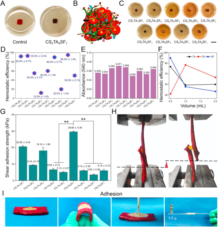
In recent years, the developed hemostatic technologies are still difficult to be applied to the hemostasis of massive arterial and visceral hemorrhage, owing to their weak hemostatic function, inferior wet tissue adhesion, and low mechanical properties. Herein, a mussel-inspired supramolecular interaction-cross-linked hydrogel with robust mechanical property (308.47 ± 29.20 kPa) and excellent hemostatic efficiency (96.5% ± 2.1%) was constructed as a hemostatic sealant. Typically, we combined chitosan (CS) with silk fibroin (SF) by cross-linking them through tannic acid (TA) to maintain the structural stability of the hydrogel, especially for wet tissue adhesion ability (shear adhesive strength = 29.66 ± 0.36 kPa). Compared with other materials reported previously, the obtained CS/TA/SF hydrogel yielded a lower amount of blood loss and shorter time to hemostasis in various arterial and visceral bleeding models, which could be ascribed to the synergistic effect of wound closure under wet state as well as intrinsic hemostatic activity of CS. As a superior hemostatic sealant, the unique hydrogel proposed in this work can be exploited to offer significant advantages in the acute wound and massive hemorrhage with the restrictive access of therapeutic moieties.

Keywords: Arterial and visceral bleeding models; Hemostatic sealant; Mussel-inspired hydrogel; Robust tissue anchor; Supramolecular cross-linking.


Conflict of interest statement

The authors declare that they have no known competing financial interests or personal relationships that could have appeared to influence the work reported in this paper.

Figures

Image 1
Graphical abstract
Scheme 1
Scheme 1
Schematic illustration of (A) preparation and (B) cross-linking-network structure of CS/TA/SF hydrogel for massive arterial and visceral hemorrhage.
Fig. 1
Fig. 1
Structural and physical properties of CS/TA/SF hydrogels with different weight ratios. (A) Photographs showing gross morphologies of hydrogels onto polypropylene dish. (B) SEM images of hydrogel after lyophilization. Right is the higher magnification image of the retangular area in the left one. (C) Tensile testing photograph of hydrogel. (D) Representative tensile stress−strain curves. (E) Comparison of tensile strength, strain, and Young's modulus of hydrogels with different weight ratios. (F) Contact angles for water droplet on hydrogel surface. (G) Conformational analysis of hydrogel by FTIR spectroscopy. (H) 1H NMR spectra of hydrogel. All statistical data are represented as mean ± SD (n = 3; ***P < 0.001).
Fig. 2
Fig. 2
Hemostatic performances and wet shear adhesion strengths of CS/TA/SF hydrogels with different weight ratios. (A) Photographs showing clot formation on hydrogel and polypropylene dish. (B) Schematic illustration of hemostatic mechanism. (C) Photographs showing blood pro-coagulant efficacies of hydrogels (scale bar = 10 mm). (D) Hemostatic efficiencies of hydrogels. (E) Absorbance of hemoglobin tested by microplate reader. (F) Changes of hemostatic efficiencies with different volume ratios of CS, TA, and SF. (G) Shear adhesion strength of hydrogel using wet porcine skin as substrate. (H) Lap shear testing photographs of hydrogel using an Instron machine 1185. (I) Photographs showing a strong adhesion of hydrogel with wet porcine skin and glass. All statistical data are represented as mean ± SD (n = 3; *P < 0.05, **P < 0.01, ***P < 0.001).
Fig. 3
Fig. 3
Comprehensive performance of CS2/TA1/SF1 hydrogel with optimized weight ratio. (A) Representative tensile stress−strain curve. (B) WCA measurement. (C) Blood clotting test and (D) BCI of control and sample groups. (E) Whole blood clotting kinetics of hydrogel. (F) Digital photographs of blood absorption kinetics test as a function of time. (G) Photographs of blood coagulation treated with CS/TA/SF hydrogel or nothing. (H) SBF and blood uptake ability of hydrogel. (I) SEM morphologies of RBC adhesion on hydrogel. Below is the higher magnification image of the retangular area in the above one. (J) CCK-8 assay result of LO2 cells. (K) Live-dead staining images of LO2 cells encapsulated in hydrogel after seven days of incubation (scale bar = 100 μm). All statistical data are represented as mean ± SD (n = 3; ***P < 0.001).
Fig. 4
Fig. 4
Hemostatic property of CS2/TA1/SF1 hydrogel in various damage models of rats. (A) Hemostatic images of covering gauze or hydrogel on various wounds with no treatment as a control (scale bar = 1 cm). (B) Comparison of bleeding area in the liver injury model. (C) Quantitative analysis of bleeding area for each group. All statistical data are represented as mean ± SD (n = 3; ***P < 0.001).
Fig. 5
Fig. 5
Hemostatic property and post-operative analysis of CS2/TA1/SF1 hydrogel in various massive hemorrhage models of rabbits. (A) Rabbit ear artery, (B) liver, (C) cardiac puncture, and (D) femoral artery injury models treated by hemostatic hydrogel (scale bar = 1 cm). (E) Photograph and H&E staining image of interface reaction between rabbit cardiac tissue and hydrogel. (F) MAP through a rabbit's carotid artery and ECG of a rabbit before and after surgery. (G) Comparison of hemostatic speed between CS2/TA1/SF1 hydrogel and previously-reported hemostatic dressings.

References

    1. Han W., Zhou B., Yang K., Xiong X., Luan S., Wang Y., Xu Z., Lei P., Luo Z., Gao J., Zhan Y., Chen G., Liang L., Wang R., Li S., Xu H. Biofilm-inspired adhesive and antibacterial hydrogel with tough tissue integration performance for sealing hemostasis and wound healing. Bioact. Mater. 2020;5(4):768–778. - PMC - PubMed
    1. Wang A.Y., Rafalko J., MacDonald M., Ming X., Kocharian R. Absorbable hemostatic aggregates. ACS Biomater. Sci. Eng. 2017;3(12):3675–3686. - PubMed
    1. Leonhardt E.E., Kang N., Hamad M.A., Wooley K.L., Elsabahy M. Absorbable hemostatic hydrogels comprising composites of sacrificial templates and honeycomb-like nanofibrous mats of chitosan. Nat. Commun. 2019;10(1):2307. - PMC - PubMed
    1. Arnaud F., Tomori T., Carr W., McKeague A., Teranishi K., Prusaczyk K., McCarron R. Exothermic reaction in zeolite hemostatic dressings: QuikClot ACS and ACS+®. Ann. Biomed. Eng. 2008;36(10):1708–1713. - PubMed
    1. Hong Y., Zhou F., Hua Y., Zhang X., Ni C., Pan D., Zhang Y., Jiang D., Yang L., Lin Q., Zou Y., Yu D., Arnot D.E., Zou X., Zhu L., Zhang S., Ouyang H. A strongly adhesive hemostatic hydrogel for the repair of arterial and heart bleeds. Nat. Commun. 2019;10(1):2060. - PMC - PubMed

LinkOut - more resources